Loading component...
Ear infection: what the owner needs to know
Written by Alberto Martín Cordero
Otitis externa can be frustrating for both the owner and the clinician, as treatment requires a lot of effort, often for a protracted period of time. This article details the minimum information that should be provided to the cat and dog owner when the problem is first identified.
Introduction
Otitis externa is a common condition in dogs and cats, with a reported incidence of 10-20% in dogs and 2-6% in cats [1],[2],[3]. Predisposing, primary, secondary and perpetuating factors should be identified wherever possible in order to control the condition. Predisposing factors include anatomical conditions such as a stenotic ear canal, excess hair within the canal, increased moisture (e.g., in certain breeds with pendulous pinnae or in dogs that swim), and over-treatment. There are various possible primary factors; the most common are skin allergies, although foreign bodies, hypersecretory conditions (e.g., primary seborrhea, hypothyroidism, or increased ceruminous gland activity), neoplasia and parasites are also common [4]. Secondary factors include bacterial and yeast infections, whilst the main perpetuating factors are otitis media and chronic pathological changes in the ear canal secondary to inflammation (e.g., stenosis, fibrosis and calcification of tissues). The correct techniques for ear examination, sampling and cleaning are key points in treating, diagnosing and managing otitis externa in dogs. The primary cause must be identified and treated, and any secondary factors must be eliminated. If there are chronic pathological changes present these must be properly controlled for satisfactory long-term management.
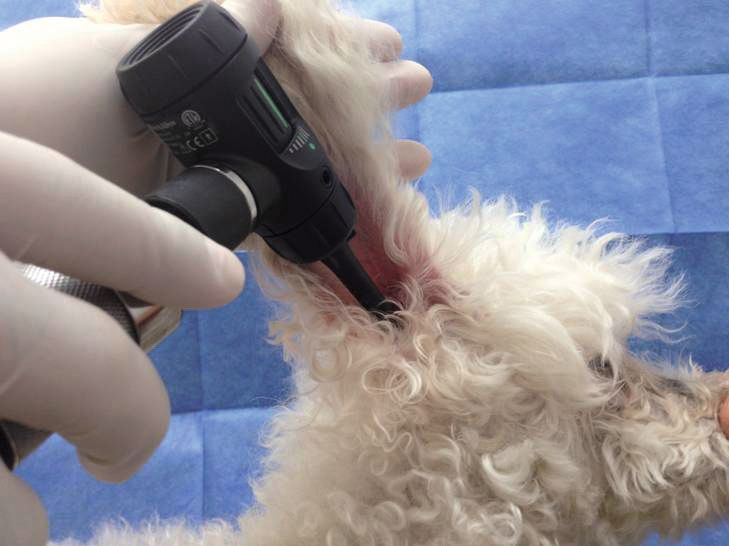
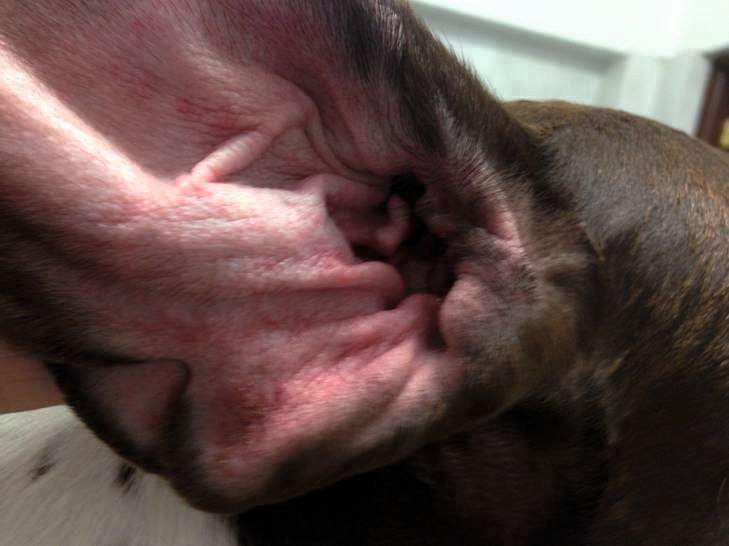
Ear examination
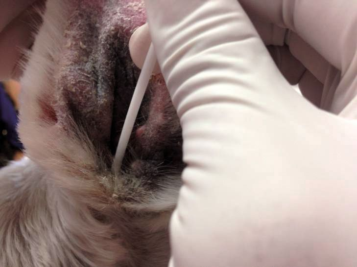
Ear cytology sampling
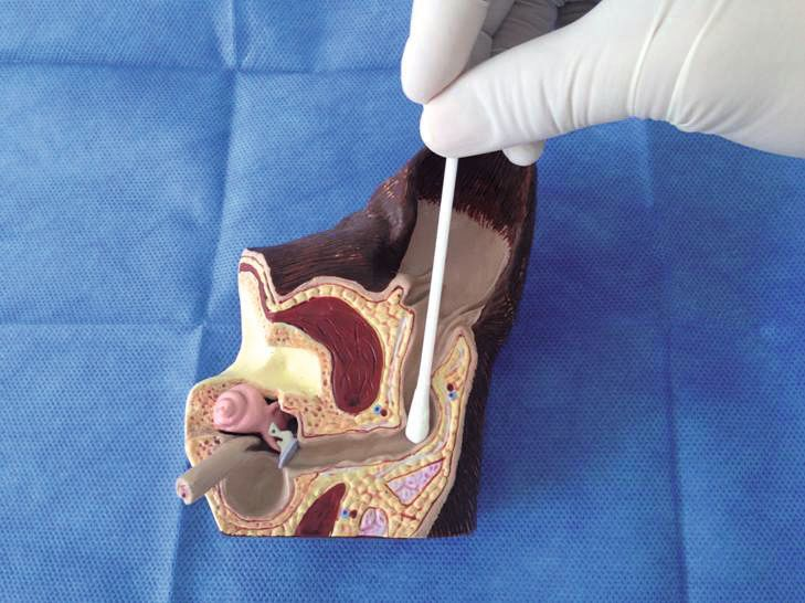
Ear cleaning
Superficial ear cleaning does not require anesthesia or sedation in the majority of patients, and owners should be instructed as to how to perform this procedure properly at home. In most cases of otitis externa, epithelial migration, the self-cleaning mechanism of the ear canal, is affected adversely, leading to cerumen accumulation [6],[7].


References
- Baba E, Fukata T, Saito M. Incidence of otitis externa in dogs and cats in Japan. Vet. Rec. 1981;108:393-395.
- Griffin CE, Song M. Otitis workshop. In: Kwochka K, Willemse T, von Tscharner C (eds). Advances in Veterinary Dermatology, vol. 3. Boston: Butterworth-Heinemann 1996;369-375.
- Rosychuk RA, Luttgen P. Diseases of the ear. In: Ettinger SJ, Feldman EC (eds.) Textbook of Veterinary Internal Medicine: Diseases of the Dog and Cat. 5th ed. Philadelphia: WB Saunders 2000;1185-1235.
- Saridomichelakis MN, Farmaki R, Leonidas LS, et al. Aetiology of canine otitis externa: a retrospective study of 100 cases. Vet. Dermatol. 2007;18:341-347.
- Campbell JJ, Coyner KS, Rankin SC, et al. Evaluation of fungal flora in normal and diseased canine ears. Vet. Dermatol. 2010;21(6);619-625.
- Tabacca NE, Cole LK, Hillier A, et al. Epithelial migration on the canine tympanic membrane. Vet. Dermatol. 2011;22(6);502-510.
- Nuttall T, Cole LK. Ear cleaning: the UK and US perspective. Vet. Dermatol. 2004;15(2):127-136.
Alberto Martín Cordero
DVM, VETDERM Dermatology Specialists, Guadalajara, Mexico
Alberto Cordero is a dermatologist and founder of “Vetderm”, a referral clinic in Guadalajara, Mexico. After graduating as a veterinarian from the University of Guadalajara, he worked at the Animal Dermatology Clinic in California, USA and at the Ludwig Maximilian University in Munich, Germany. He received a diploma in dermatology from the University of Luxembourg’s European School for Advanced Veterinary Studies (ESAVS). A member of the American Academy of Veterinary Dermatology, he is also part of the Programming Committee for the North American Veterinary Dermatology Forum (NAVDF), and a founding member of the Latin American Society of Veterinary Dermatology (SLDV). Dr. Cordero lectures at numerous congresses in Mexico, Central and South America, and Europe.
Other articles in this issue
Share on social media