Loading component...
The three most common oral pathologies in adult cats
Written by Javier Collados
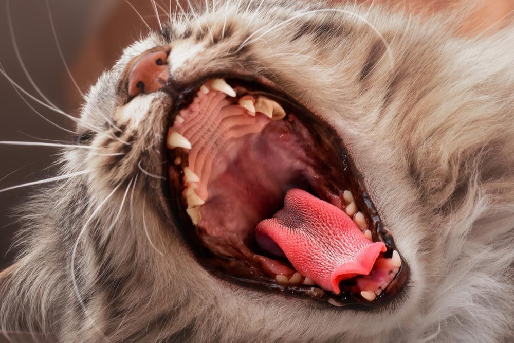
The first step in detecting oral disease is to perform an initial oral examination in the awake animal. However, in order to thoroughly detect disease a complete oral examination must be performed under general anesthesia.
© Shutterstock
Key points
A dental explorer, periodontal probe and intraoral radiography are essential tools for the diagnosis and staging of most dental problems.
The most commonly diagnosed oral diseases in adult cats are chronic periodontitis, dental fractures, and tooth resorption.
Periodontal disease
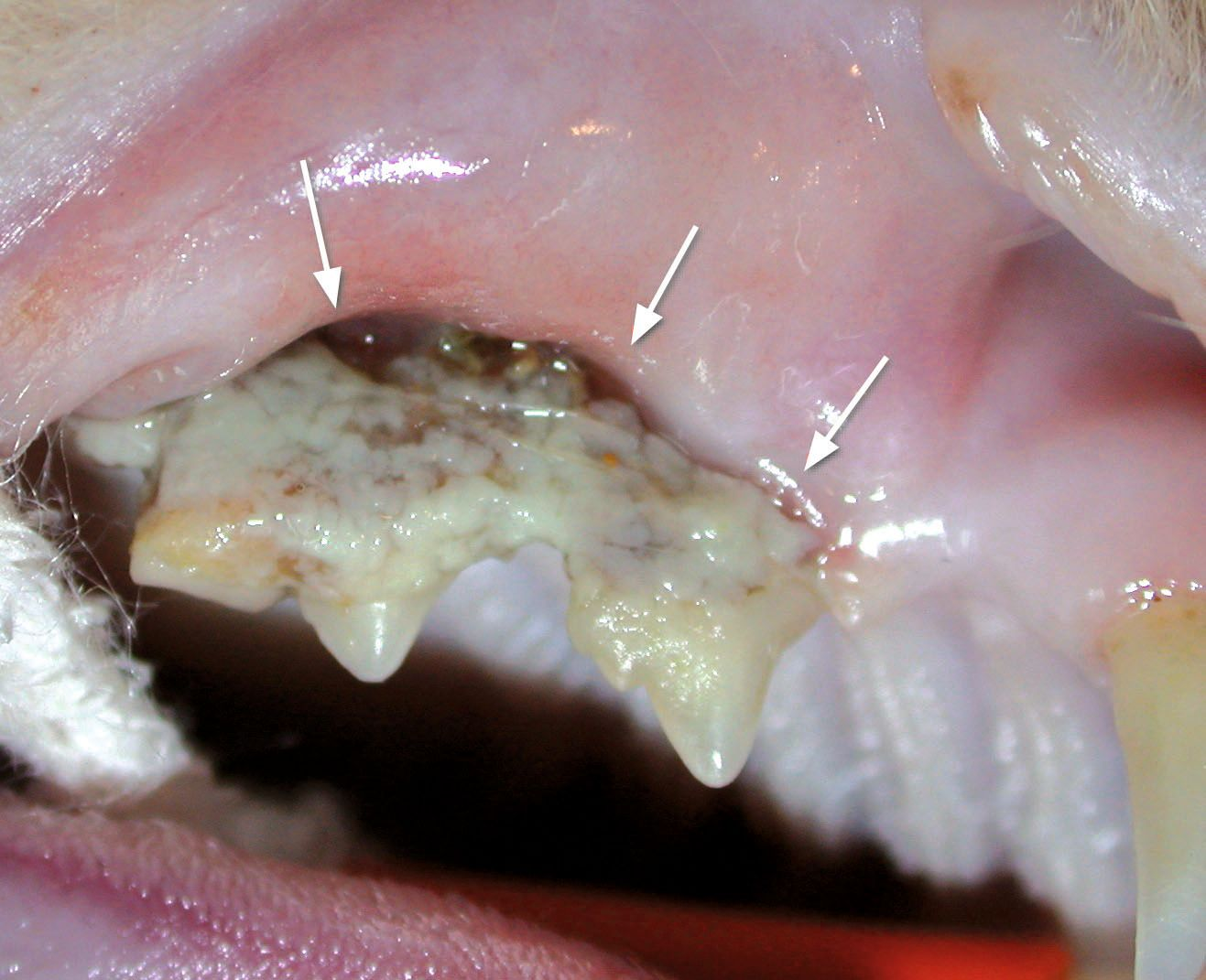
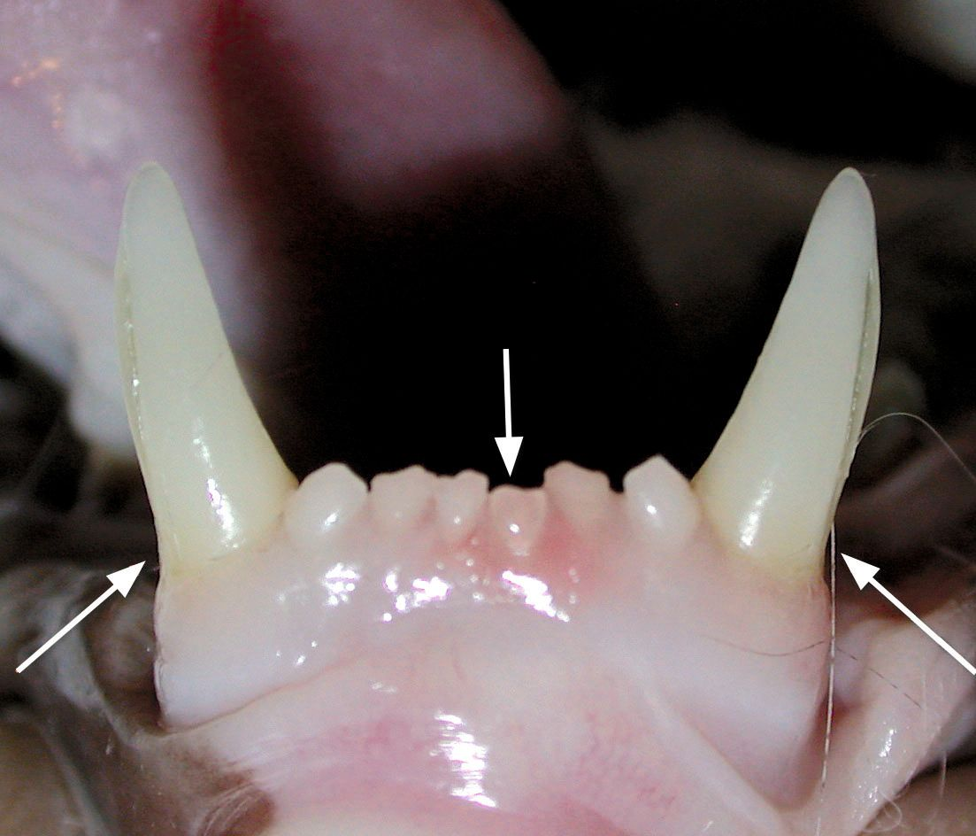
Advanced periodontal disease is commonly diagnosed in cats (Figure 1). A major contributing factor to its development is the lack of adequate oral hygiene at home. The adoption of preventive pediatric health plans and adequate geriatric presurgical profiles, as well as the presence of specialists in oral surgery and anesthesia, are crucial aspects for ensuring appropriate periodontal treatment in such patients. Staging of periodontal disease is essential for the decision-making process, which can range from calculus removal and dental polishing to surgical extraction.
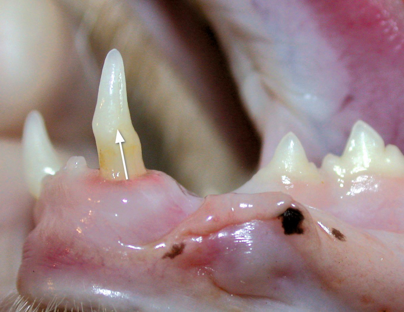
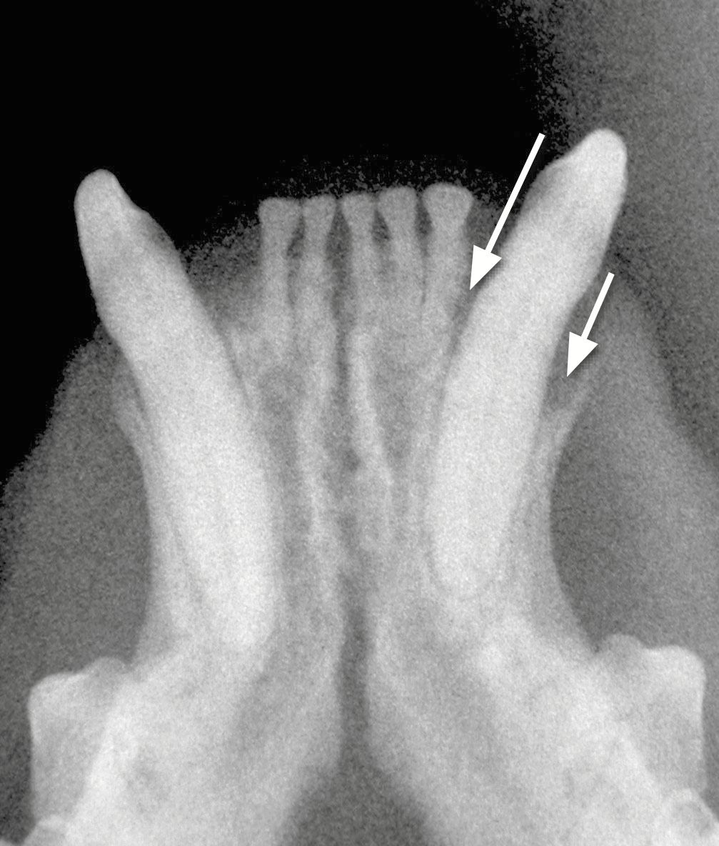
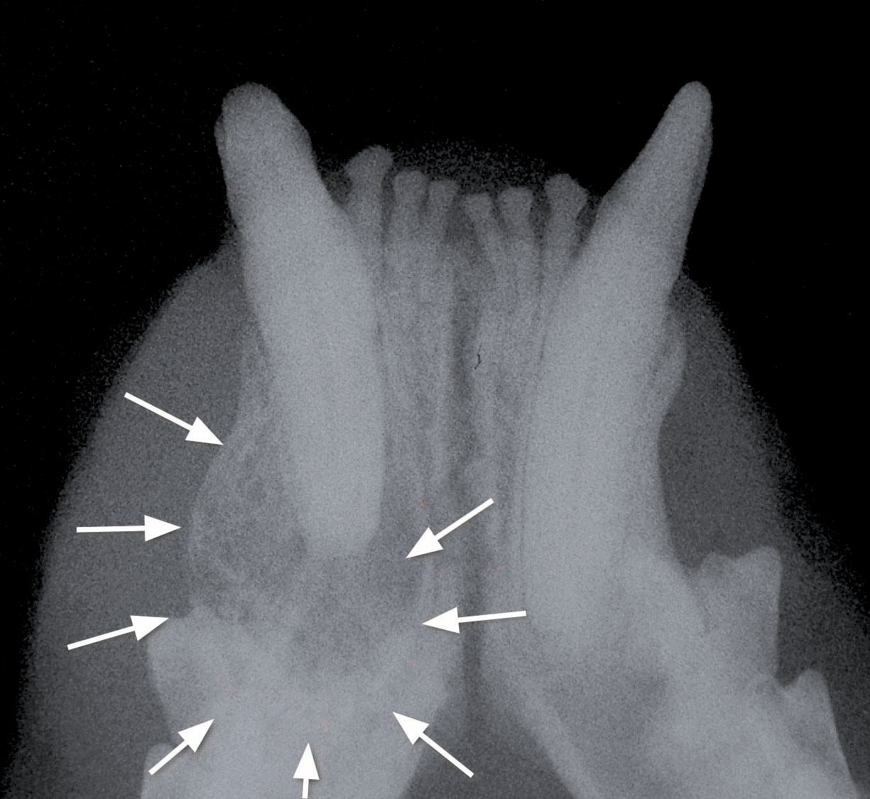
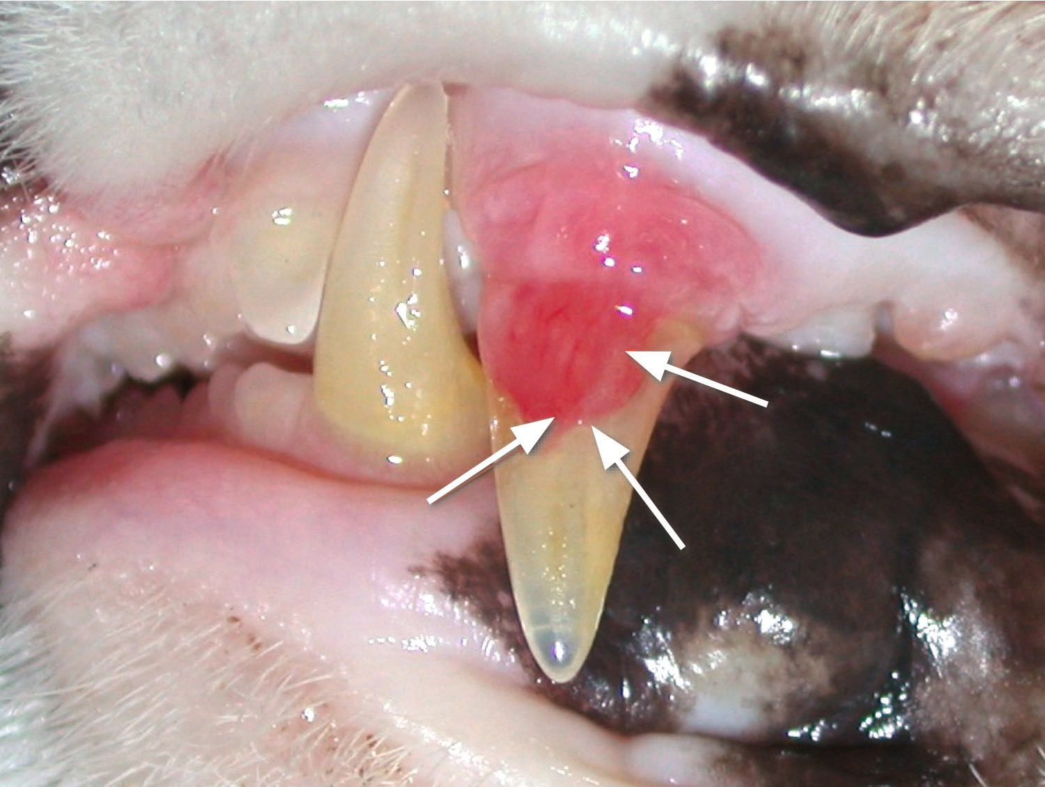
Tooth extrusion (Figure 2) is a sign of advanced periodontal disease in cats. Adequate periodontal probing and intraoral radiography (Figure 3) are very important when staging these teeth, as they are essential components in the decision-making process for managing the condition.
Dental fractures
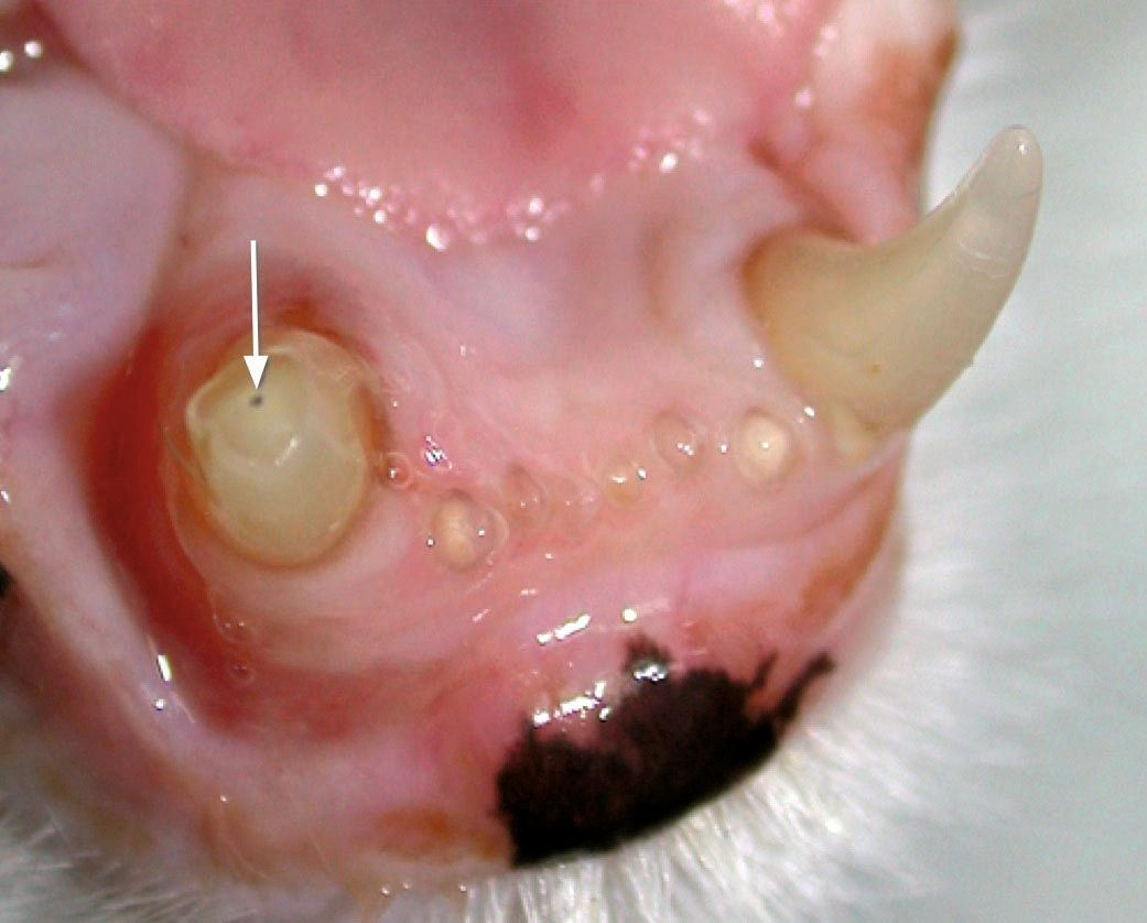
A tooth fracture is characterized by structural alteration (and in most cases loss) of dental tissue secondary to external trauma to the oral cavity. Note that fractures are frequently missed on the initial oral examination performed on a conscious animal. As in periodontal disease, classification is essential for decision making. This is particularly important in adult patients, since fractures involving exposure of the pulp chamber or cavity (complicated fractures, root fractures) that have not been adequately treated can lead to clear signs of pulp disease, such as dental abscesses, fistulas, etc. (Figure 4 and 5).
Tooth resorption
Tooth resorption (TR) is a primary dental disease characterized by progressive tissue destruction of one or more permanent teeth due to the action of odontoclastic cells. The condition frequently manifests with resorption of the crown and/or neck of the tooth, along with reactive gingival hyperplasia (Figure 6).
The underlying cause is complex and has not been clearly established. Although TR is not unique to adult animals, its progression in different stages and the appearance of obvious signs in the oral cavity are usually observed in adult animals. Nevertheless, radiography is essential for the diagnosis and treatment of TR in cats.
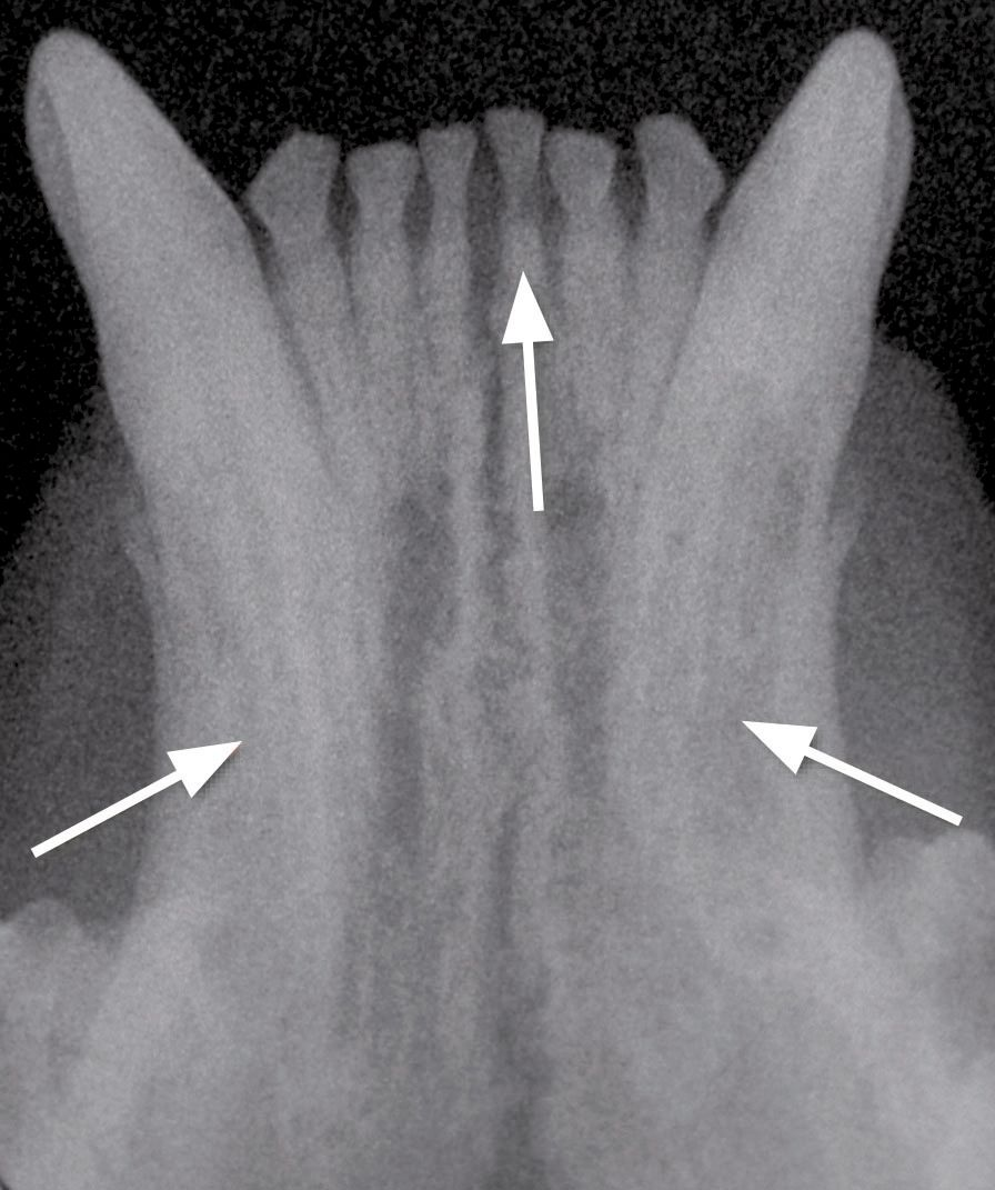
In some cases, there may be no indication that the crown of the tooth is affected, despite radiological evidence of severe root resorption (Figure 7 and 8).
Note teeth are identified using the American Veterinary Dental College (AVDC) classification system.
Acknowledgement: many thanks to Dr. Carlos Rice for reviewing the English version of the original Spanish article.
Javier Collados
DVM, PhD, MRCVS
Spain
Dr. Collados graduated in Veterinary Medicine from the Complutense University of Madrid in 1994 and earned a PhD from the same university in 2021. He completed a residency at the American Veterinary Dental College (AVDC). He was the only AVEPA certified in Dentistry and Oral Surgery in 2013, working exclusively in this field. He heads the Dentistry and Oral Surgery service at Sinergia Veterinaria. He was a lecturer and subject coordinator in Animal Dentistry at the Faculty of Veterinary Medicine of the Alfonso X el Sabio University of Madrid.
He has authored numerous publications and, more particularly, the Visual Atlas of Oral and Dental Pathologies in Exotic and Small Animals (Ed. Servet, 2008), translated into Spanish, French and Japanese. He has spoken at national and international courses and congresses, and has participated in more than 100 events.
References
- https://avdc.org/avdc-nomenclature/ (downloaded May 15, 2014).
Share on social media