Anesthesia for cesarean section in the dog
Written by Bonnie Hay Kraus
The major goal in anesthesia for cesarean section (CS) is to minimize fetal effects of anesthetic drugs in order to minimize fetal respiratory, central nervous system and cardiovascular depression and deliver live, vigorous puppies.
Article

Key points
The major goals for cesarean section are to deliver live, vigorous puppies while providing adequate analgesia to the dam.
There is a higher anesthetic risk with cesarean section due to pregnancy-associated physiologic changes.
Planning and preparation are important for both elective and emergent surgical scenarios.
Optimized ventilation, oxygenation and perfusion in the dam should permit a “happy dam, happy baby” scenario.
Neonatal resuscitation centers on stimulating respiration and supporting oxygenation and body temperature.
Most commonly used analgesic drugs may be safely administered to lactating dams without adversely affecting the neonates.
Introduction
The major goal in anesthesia for cesarean section (CS) is to minimize fetal effects of anesthetic drugs in order to minimize fetal respiratory, central nervous system and cardiovascular depression and deliver live, vigorous puppies. Of equal importance is to provide adequate analgesia to the dam and prevent anesthesia-related complications such as hypotension, hypoventilation, hypoxemia, hemorrhage and hypothermia, which will increase morbidity and mortality in both mother and puppies. The physiochemical properties which allow drugs to cross the blood-brain barrier also facilitate crossing of the placenta, therefore the assumption should be made (with very few exceptions) that anesthetics, analgesics and sedatives/tranquilizers all cross the placenta. Prolonged labor prior to delivery causes maternal physiologic compromise, resulting in fetal depression due to decreased placental perfusion, hypoxemia and acidosis. Maternal and puppy mortality is significantly increased during emergent versus planned CS [1] [2]. Timing and preparation are extremely important for puppy survival for both elective and emergency CS, and a thorough understanding of the maternal physiologic changes and the potential impact of anesthetic drugs is essential to optimize outcomes for both mother and fetus (Figure 1).

Maternal physiologic changes
Increased metabolic demands imposed by the fetus result in major physiologic changes during pregnancy and impact the anesthetic management of these patients (Figure 2). Much of the data describing these alterations have been obtained in humans and sheep, but should be comparable – if not greater in magnitude – in the dog, since birth weight as a percentage of maternal weight is significantly greater [3]. Pregnancy-associated physiologic changes to the cardiac, pulmonary and gastrointestinal (GI) systems are summarized in Table 1. These result in greater anesthetic risk (due to decreased cardiac and respiratory reserve and a higher chance of vomiting/regurgitation with aspiration) as well as decreased anesthetic requirements (which may put them at risk for anesthetic overdose) [3] [4].| Cardiovascular |
↑Heart rate, stroke volume, cardiac output
↓Vascular tone, arterial blood pressure
↑Oxygen consumption
↑Red blood cells, blood/plasma volume
↓Packed cell volume/hemoglobin/plasma proteins
|
| Respiratory |
↑Respiratory rate, tidal volume, minute ventilation
↓Functional residual capacity
|
| Gastrointestinal |
↓Lower esophageal sphincter tone
↑Intra-gastric pressure/gastric emptying time
↓GI motility, pH of gastric secretions
↑Gastrin production
|
| Central Nervous System | ↑Endorphins |

Cardiovascular
The growing fetuses increase metabolic demand and maternal oxygen consumption. Increases in heart rate and stroke volume augment cardiac output by 30-40% to meet the demand [3] [4] [5], but result in decreased cardiac reserve.
Unlike other major organs, uterine blood flow is not auto-regulated [4]. Uterine blood flow and placental perfusion are directly related to systemic blood pressure and inversely proportional to myometrial vascular resistance [3]. Decreases in uterine blood flow will result in decreased fetal oxygen delivery. Pain, stress, hyperventilation and some drugs (e.g., alpha-2 agonists) can all decrease cardiac output during labor and contribute to decreased uterine blood flow. Control of pain and anxiety are key components of successful patient management. Care must be taken to avoid cardiac depression from excessive doses of sedatives or anesthetics. In humans, the posterior vena cava and aorta can be compressed during dorsal recumbency which decreases venous return, cardiac output and uterine blood flow. Although less significant in dogs, the amount of time in dorsal recumbency should minimized [3] [4].
Maternal blood volume during pregnancy increases by up to ~23% in dogs, along with red blood cells (RBCs) [6], with plasma volume increasing more than RBCs, leading to decreased packed cell volume. This pregnancy-associated anemia increases in magnitude in relation to the number of fetuses [7]. Increased blood volume buffers against losses during parturition but can confound the use of packed cell volume as a measure of dehydration preoperatively; other clinical signs may need to be utilized. There is a greater risk of intra-operative hemorrhage due to the increased blood flow to the gravid uterus (20-40 times normal) and mammary glands [5]. Intra-operative hemorrhage should be quantified and replaced with 3-4 times the amount in crystalloid solution (up to 10% loss of the total blood volume) to avoid the associated hypotension and decreased uterine blood flow. Colloid therapy should be added if hemorrhage reaches 20%. Hypotension may be treated with ephedrine (administered as a bolus, 0.03-0.1 mg/kg IV); this is the drug of choice in pregnant women since it increases blood pressure while maintaining uterine blood flow, whereas both dopamine and dobutamine decrease uterine blood flow [3] [4].
Pulmonary
Tidal volume, respiratory rate and minute ventilation all increase, but functional residual capacity (FRC) decreases due to craniodorsal displacement of the abdominal organs and diaphragm by the gravid uterus [3] [4] [5]. Decreased FRC leads to small airway closure and atelectasis. The combination of decreased FRC and increased oxygen consumption raises the risk of hypoxemia during periods of hypoventilation or apnea (e.g., during anesthetic induction [3] [4]). Pre-oxygenation prior to anesthetic induction is recommended to delay the onset of hypoxemia from approximately 60 seconds to up to five minutes if the patient is tolerant [8].
Increased serum progesterone decreases lower esophageal sphincter tone, GI motility and gastric emptying, whilst cranial displacement of the stomach increases intra-gastric pressure; together these contribute to a greater risk of regurgitation and aspiration [3] [4]. Increased gastrin and gastric acid production lowers stomach pH and puts parturient patients at greater risk for aspiration pneumonitis and esophagitis [4]. Prophylactic administration of metoclopramide or anti-emetics such as maropitant or ondansetron and/or H2 receptor antagonists may help ameliorate these effects. Emergent patients are also likely to be inadequately fasted, and rapid IV induction followed by intubation (ensuring the cuff is properly inflated) is recommended.
CNS
General pharmacology and pregnancy
Placental transfer of drugs has been primarily studied in sheep and laboratory animals, and direct extrapolation to dogs may be misleading due to species differences in placentation, extent of placental metabolism and transport of drug across the placenta [4]. However, in general, the same physiochemical properties that allow drugs to cross the blood brain barrier also facilitate their placental transfer. The safest assumption is that most, if not all, drugs cross the placenta and affect the fetus. Elective procedures requiring anesthesia are best avoided during the first trimester (20 days of gestation in dogs), when the fetuses are most vulnerable to teratogenic effects of drugs.
Simple diffusion is the most important mechanism of placental transfer of drugs. Properties favoring transfer include:
- Molecular weight < 600 Da
- High lipid solubility
- Low degree of protein binding
- Non-ionization at maternal blood pH [3] [4]
Most anesthetic drugs, with the exception of glycopyrrolate and neuromuscular blocking agents, have MW < 300 Da and are relatively lipid soluble, and thus readily cross the placenta.
Drug protein binding and degree of ionization is determined by its pKa and blood pH, which in turn can affect the distribution between the dam and fetus. As blood pH decreases, acidic drugs such as thiobarbiturates will be less ionized and the fraction of drug bound to protein is decreased, leading to greater clinical effect [3] [4]. Weakly basic drugs (opioids, local anesthetics) become more highly ionized, resulting in less effect on the dam and fetus [3] [4]. Redistribution of drugs out of the fetus back into the dam’s circulation as maternal plasma levels decrease makes clinical estimation of fetal plasma concentrations difficult. Although ~50% of umbilical vein blood passes through the fetal liver, microsomal enzyme activity and metabolism are minimal [4].
Inhalant agents readily cross the placenta and should be titrated to the lowest level needed to achieve adequate anesthesia. Unlike halothane and methoxyflurane, which rely heavily on metabolism (~20-50% and 50-75% respectively) for elimination, isoflurane and sevoflurane are almost entirely eliminated through the respiratory system. Additionally, the low blood solubility results in rapid clearance from the newborn as long as they are breathing at delivery. It is important to avoid high inhalant concentrations to prevent neonatal respiratory depression and apnea. Isoflurane is associated with improved puppy survival at 7 days compared to methoxyflurane and there is no difference when compared to epidural anesthesia [1].
- Anticholinergic agents such as atropine and glycopyrrolate are primarily used to decrease vagal tone due to opioids or traction on the uterus, or to support fetal heart rate. Choice depends on the desire for placental transfer, since atropine crosses the placental barrier but glycopyrrolate does not. Glycopyrrolate will mitigate the increased vagal tone caused by mu agonist opioids and prevent maternal bradycardia and possible hypotension. It also increases gastric pH and may decrease the severity of chemical pneumonitis should regurgitation and aspiration occur in the dam [3]. Fetal bradycardia (< 150 beats/min) indicates fetal distress and is one of the prime indicators for emergency CS [10]. Fetal cardiac output is more dependent on heart rate than blood pressure. Atropine may be administered to the dam if the objective is to increase fetal heart rate, which may be low due to hypoxemia, fetal distress or mu agonist opioids. Increasing the fetal heart rate will increase myocardial oxygen consumption in the face of hypoxemia, which may lead to myocardial ischemia, so atropine use is controversial. However, it may support the heart rate long enough to allow delivery and proper resuscitation. It is important to optimize maternal oxygenation, cardiac output and blood pressure, and ensure good ventilation for the puppies after delivery.
- Tranquilizers/sedatives are typically avoided due to cardiorespiratory and CNS depression. Xylazine alone or in combination with ketamine is associated with higher fetal death [1]. Medetomidine at low doses (< 20 µg/kg) does not increase uterine muscle activity or cause abortion [3], but both medetomidine and dexmedetomidine cause significant decreased cardiac output in the dam due to vasoconstriction and baroreceptor-mediated bradycardia, and drug label recommendations advise against their use in pregnant dogs. Benzodiazepines (diazepam and midazolam) can cause neonatal depression, lethargy, apnea and hypothermia, especially at higher doses. Low dose acepromazine may be an option for very stressed or anxious dogs to avoid decreased uterine blood flow. The drug crosses the placenta slowly due to its higher molecular weight and protein binding, and is not associated with increased maternal or neonatal mortality [2] [4], but its α-adrenergic antagonism can cause vasodilation and therefore it should be avoided in dehydrated or compromised patients.
- Opioids include mu-receptor agonists such as morphine, hydromorphone, oxymorphone, fentanyl, methadone and meperidine. Buprenorphine is a partial muagonist and butorphanol is a mu-receptor antagonist and a kappa agonist. These latter two drugs typically produce less sedation and respiratory depression than full mu agonists, but offer less potent analgesia.
Administering opioids to the dam will result in placental transfer, the extent of transfer differing depending on the drug. Less than 10% of buprenorphine is transferred to the fetus, whereas fentanyl (which is highly lipid soluble) crosses the placenta in high amounts, persisting long after maternal clearance [4]. Of the commonly used mu agonists, morphine is the least lipid soluble and only 20-30% is non-ionized at normal plasma pH [4]. It crosses the placenta less rapidly than more lipid soluble agonists such as fentanyl. The lipid solubility of hydromorphone lies between these two drugs. Neonates are significantly more sensitive to CNS and respiratory depression of opioids due to the immaturity of their CNS, increased permeability of the blood-brain barrier and end-organ sensitivity, and even small changes in ventilation may lead to hypoxemia and increased mortality. Respiratory and CNS depression in newly delivered neonates can be reversed with naloxone
.
Anesthetic induction
The use of thiopental, ketamine, xylazine and methoxyflurane have been associated with increased puppy mortality and/or decreased puppy vigor at birth, and are best avoided [1] [11] [12]. Epidural anesthesia/analgesia used alone for CS has minimal fetal effects but has various drawbacks; it is technically difficult, airway protection via intubation is impossible, and there is the risk of hind-limb paralysis, hypotension or urinary retention (if epidural opioids are used).
Propofol has a short half-life and rapid metabolism, including extra-hepatic metabolism, but it can cause cardiopulmonary depression depending on the dose and rate of administration. Propofol followed by isoflurane has puppy survival rates equivalent to epidural anesthesia and is associated with a positive effect on neonatal survival at 7 days [1]. Propofol has similar fetal mortality rates to mask induction with isoflurane, but it allows IV induction and rapid control and protection of the airway [1] [13].
Alphaxalone is available in many countries and has been shown to have a shorter terminal half-life compared to propofol [14] [15]. Two recent studies comparing propofol to alphaxalone for induction at CS found no significant difference in puppy mortality at 24 hours or up to 3 months after delivery, but both studies identified differences in “puppy vigor”. Apgar scores and all four health vigor assessments (withdrawal, sucking, anogenital and flexion reflexes) were greater for puppies for up to 60 minutes post-delivery where the dams received alphaxalone [16] [17].
Anesthetic technique
As noted above, the increased risk for regurgitation and aspiration can be decreased using maropitant (1.0 mg/kg SC administered at least 30 minutes prior to opioid administration [18]). For more emergent situations, maropitant may be given (1.0 mg/kg IV over 5 minutes while monitoring blood pressure) and discomfort on injection can be decreased by diluting 50:50 with crystalloid solution. Alternatively, an opioid premedication which does not induce vomiting may be administered (e.g., butorphanol at 0.2-0.3 mg/kg IM or IV). Sedatives, e.g., acepromazine at 2.0-5.0 µg/kg IM or IV, should be reserved for highly stressed dams; however, remember that this drug has a long duration of action and cannot be reversed in either the dam or puppies.
Lumbosacral epidural with either local anesthetic or opioid (alone or in combination) can be administered either before or directly after induction to provide anesthesia and analgesia. Lidocaine (2.0-3.0 mg/kg, 0.1-0.15 mL/kg of 20 mg/mL concentration) will last for ~90 minutes, whilst bupivacaine (0.75-1.5 mg/kg, 0.1-0.2 mL/kg of 7.5 mg/mL concentration) lasts up to 4-6 hours. Both drugs will affect hindlimb motor function and may contribute to intra-operative hypotension through sympathetic blockade. Alternatively, preservative-free morphine may be used alone to provide analgesia (0.1-0.2 mg/kg); the onset of action may take up to 60 minutes, but motor function is not affected. Epidural opioids may cause urinary retention and – since early hospital discharge is advantageous for healthy dam and puppies – owners must monitor urine output for 24 hours after discharge. The combination of lidocaine (2.0 mg/kg) and morphine (0.1 mg/kg) provides rapid onset of anesthesia along with synergistic and long-lasting analgesia. Epidural drug administration requires much lower doses than the same drugs given parenterally and therefore decreases systemic effects in the dam and puppies. Alternatively, the epidural can be performed after the pups are delivered and the incision is closed to provide post-operative analgesia.
If local anesthetic epidural is not performed, a lidocaine block along the line of incision (at 2 mg/kg, diluted with sterile water if necessary to increase the volume) will provide intra-operative analgesia. Bupivacaine may be used (1.5-2.0 mg/kg) for an incisional block at the time of closure of the linea alba for longer post-operative analgesia. Lidocaine and bupivacaine mixtures have a decreased duration of action and are not recommended [19].
Excretion of lidocaine and bupivacaine and/or their metabolites does occur but there is minimal effect on human neonates [22] and milk concentrations of ropivacaine are lower than for other local anesthetics [23]. Although species differences may occur, based on current evidence it seems that most commonly used analgesics may be safely administered to lactating dams without adversely affecting the neonates.
Neonate resuscitation
- Laryngoscope with small blade (size 0-1)
- Endotracheal tubes (size 2.0-3.0 mm OD, 14G or 18G IV catheter)
- Naloxone
- Doxapram
- Epinephrine
|
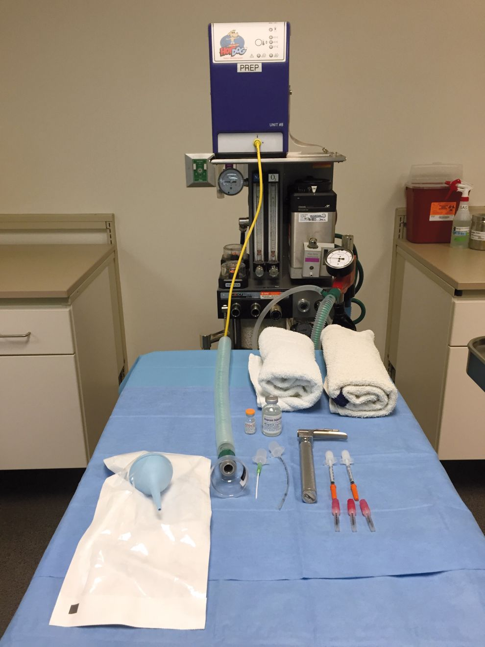
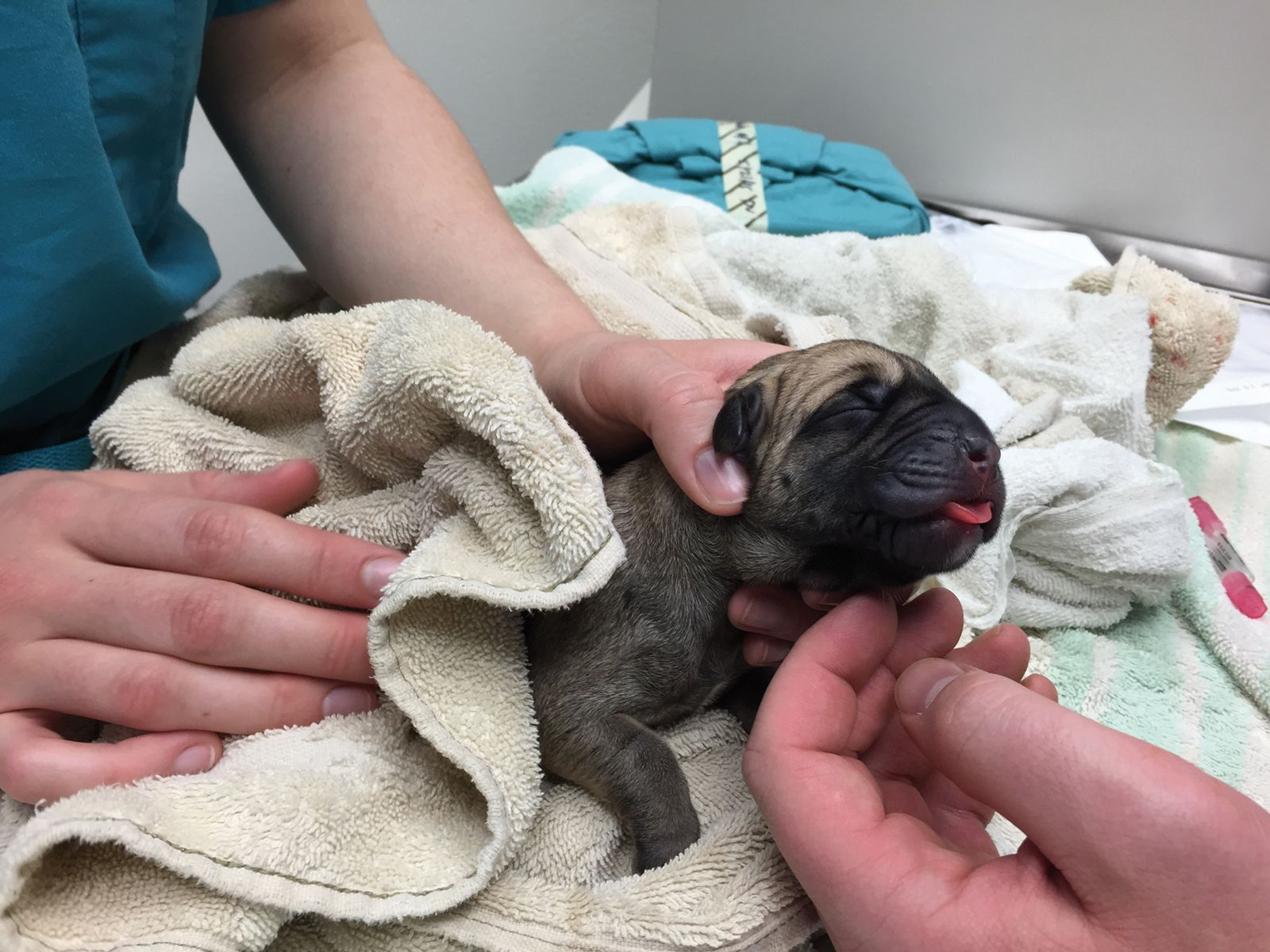
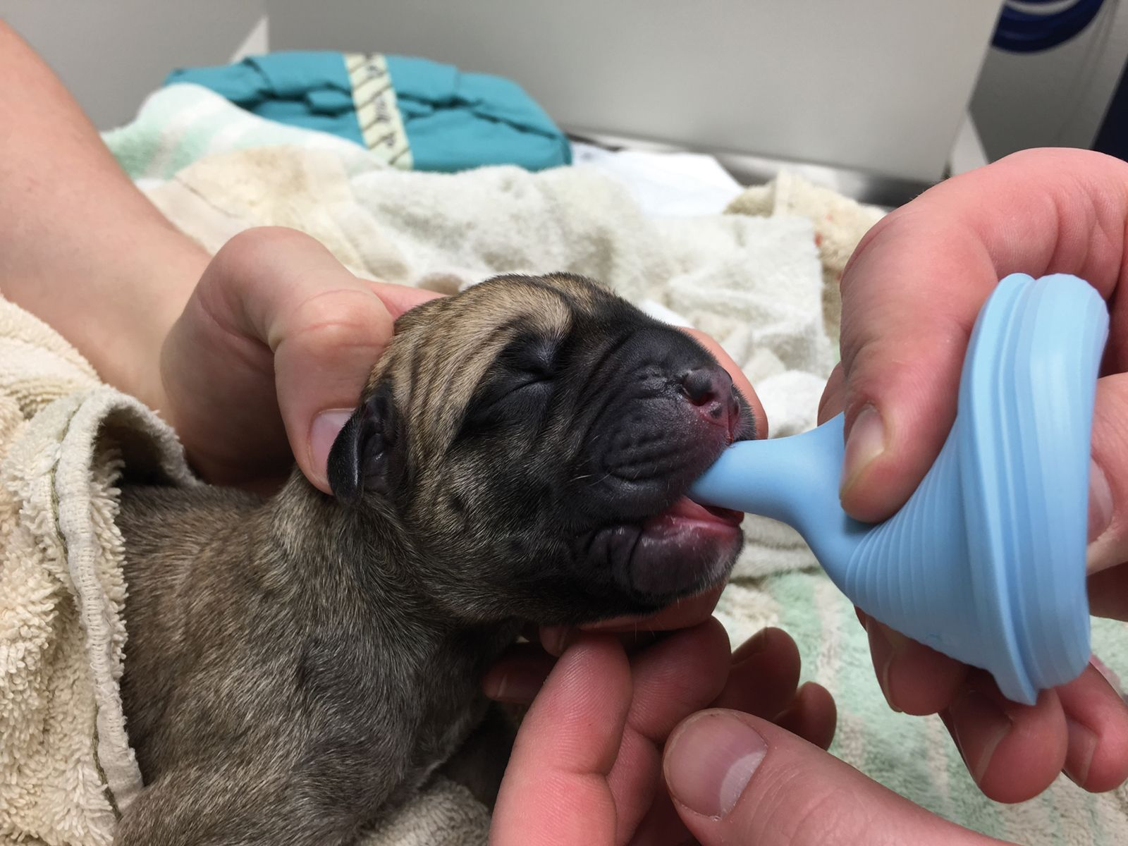

Spontaneous breathing should be identified by observing the chest wall, listening for vocalization, or auscultation with a stethoscope. The two major causes of fetal depression are hypoxemia and drugs administered to the dam. Vigorous rubbing should continue along with oxygen supplementation and gentle chest compressions. Opioids administered to the dam should be reversed in the neonate with naloxone (0.002-0.02 mg/kg IV or 1-2 drops sublingually) after delivery if the pups are slow to begin breathing, moving and vocalizing. Neonatal heart rates should be ~220 bpm and can be counted by palpating a precordial pulse. Bradycardia usually indicates hypoxemia and should be treated by stimulating ventilation, supplemental oxygen, patient warming and mechanical stimulation as described above. Doxapram (1-2 drops sublingually) can be used as a respiratory stimulant, but it also increases cerebral oxygen consumption and should only be used with oxygen supplementation. There is no evidence against employing the drug in dogs, but its use has been discontinued in humans. If spontaneous breathing is still not observed, the pup should be intubated using a short-bladed laryngoscope and (depending on the size/breed) a flexible 14 or 18G IV catheter or 2.0-3.0 OD endotracheal tube; careful handling is necessary to avoid traumatizing the delicate neonatal tissues. Severe bradycardia or asystole may be treated with epinephrine (0.1 µg/kg) diluted with 0.5 mL of crystalloid fluid, and administered though the umbilical vein (identified as the thin-walled vessel within the umbilical stump; umbilical arteries have thicker walls). Thermoregulation reflexes are underdeveloped in neonates, therefore as soon as the neonate is breathing, moving and vocalizing, they should be placed in a warmed incubator.
Bonnie Hay Kraus
DVM, Dip. ACVS, Dip. ACVAA
Dr. Hay Kraus received her DVM degree from the University of Missouri – Columbia in 1989. She completed an internship at the New Jersey Equine Clinic in Clarksburg before undertaking residencies in both Equine Surgery and Comparative Anesthesia at Tufts University School of Veterinary Medicine, completing them in 1993 and 1998 respectively. She is board certified by both the American College of Veterinary Surgeons and the American College of Veterinary Anesthesiology and Analgesia. In 2007, she joined the veterinary faculty at the Department of Veterinary Clinical Sciences, Iowa State University – College of Veterinary Medicine, where she is currently an Anesthesiologist.
References
- Moon PF, Erb HN, Ludders JW, et al. Peri-operative risk factors for puppies delivered by cesarean section in the United States and Canada. J Am Anim Hosp Assoc 2000;36:359-368.
- Smith FO. Guide to emergency interception during parturition in the dog and cat. Vet Clin Small Anim 2012;42:489-499.
- Luna SPL, Cassu RD, Castro GB, et al. Effects of four anaesthetic protocols on the neurological and cardiorespiratory variable of puppies born by caesarean section. Vet Rec 2004;154:387-389.
- Funkquist PME, Nyman GC, Lofgren AJ, et al. Use of propofol-isoflurane as an anesthetic regimen for cesarean section in dogs. J Am Vet Med Assoc 1997:211:313-317.
- Moon-Massat PF, Erb HN. Perioperative factors associated with puppy vigor after delivery by cesarean section. J Am Anim Hosp Assoc 2002;38:90-96.
- Ferre PJ, Pasloske K, Whittem T, et al. Plasma pharmacokinetics of alfaxalone in dogs after an intravenous bolus of Alfaxan-CD RTU. Vet Anaesth Analg 2006;33:229-236.
- Zoran DL, Riedesel DH, Dyer DC. Pharmacokinetics of propofol in mixed-breed dogs and greyhounds. Am J Vet Res 1993;54:755-760.
- Doebeli A, Michel E, Bettschart R, et al. Apgar score after induction of anesthesia for canine cesarean section with alfaxalone versus propofol. Theriogenology 2013:80:850-854.
- Metcalfe S, Hulands-Nave A, Bell M, et al. Multicentre, randomised clinical trial evaluating the efficacy and safety of alfaxalone administered to bitches for induction of anaesthesia prior to caesarean section. Aust Vet J 2014:92:333- 338.
- Hay Kraus BL. Effect of dosing interval on efficacy of maropitant for prevention of hydromorphone-induced vomiting and signs of nausea in dogs. J Am Vet Med Assoc 2014;245(9):1015-1020.
- Lizarraga I, Janovyak E, Beths T. Comparing lidocaine, bupivacaine and a lidocaine-bupivacaine mixture as a metacarpal block in sheep. Vet J 2013;197(2):515-518.
- Moon PF, Erb HN, Ludders JW, et al. Peri-operative management and mortality rates of dogs undergoing cesarean section in the United States and Canada. J Am Vet Med Assoc 1998;213:365-369.
- Seaton S, Reeves M and Mclean S. Oxycodone as a component of multimodal analgesia for lactating mothers after caesarean section: Relationships between maternal plasma, breast milk and neonatal plasma levels. Aust N Z J Obstet Gynaecol 2007;47:181-185.
- Ludwig B, Jordan JC, Rehm WF, et al. Carprofen in veterinary medicine. I. Plasma disposition, milk excretion and tolerance in milk-producing cows. Schweiz Arch Tierheilk 1989;131(2):99-106.
- Ortega D, Viviand X, Loree AM, et al. Excretion of lidocaine and bupivacaine in breast milk following epidural anesthesia for cesarean delivery. Acta Anaesthesiol Scand 1999;43:394-397.
- Matsota PK, Markantonis SL, Fousteri MZ, et al. Excretion of ropivacaine in breast milk during patient-controlled epidural analgesia after cesarean delivery. Reg Anesth Pain Med 2009;34(2):126-129.
- Grundy S, Liu S, Davidson A. Intracranial trauma in a dog due to being “swung” at birth. Top Comp Anim Med 2009;24:100-103.
- Raffe MR. Anesthetic considerations during pregnancy and for the newborn. In: Grimm KA, Lamont LA, Tranquilli WJ, et al (eds). Veterinary Anesthesia and Analgesia: The Fifth Edition of Lumb and Jones. Ames, IA: Wiley Blackwell, 2015;708-719.
- Aarnes TK, Bednarski RM. Cesarean section and pregnancy. In: Snyder LBC and Johnson RA (eds.) Canine and Feline Anesthesia and Co-Existing Disease. 1st ed. Ames, IA: Wiley Blackwell, 2015;299-309.
- Camann W, Ostheimer G. Physiological adaptations during pregnancy. Intern Anesthesiol Clin 1990;28:2-10.
- Brooks V, Keil L. Hemorrhage decreases arterial pressure sooner in pregnant compared with non-pregnant dogs: role of baroreflex. Am J Physiol 1994;266:1610-1619.
- Kaneko M, Nakayama H, Igarashi N, et al. Relationship between the number of fetuses and the blood constituents of Beagles in late pregnancy. J Vet Med Sci 1993;55:681-682.
- McNally EM, Robertson SA, Pablo LS. Comparison of time to desaturation between pre-oxygenated and non pre-oxygenated dogs following sedation with acepromazine maleate and morphine and induction of anesthesia with propofol. Am J Vet Res 2009;70(11):1333-1338.
- Wilson DV, Evans AT. The effect of topical treatment on esophageal pH during acid reflux in dogs. Vet Anaesth Analg 2007;34(5):339-343.
Share on social media