Introduction
For proper interpretation of hematology data in the veterinary clinic, the results must reflect the patient’s real condition in order to contribute to an accurate diagnosis and appropriate treatment. Several crucial aspects must be taken into account to ensure the quality and reliability of the results obtained on hematological analysis, and the veterinarian must be aware of these. They include the degree of quality controls used for the hematological analyzer, the necessity to identify possible sources of error (pre-analytical, analytical and post-analytical), and the need to study blood smears. The objective of this article, in question-and-answer format, is to address the most common potential pitfalls that exist when performing hematology and interpreting the results from a canine patient.
How does lipemia or hemolysis affect hematology results?
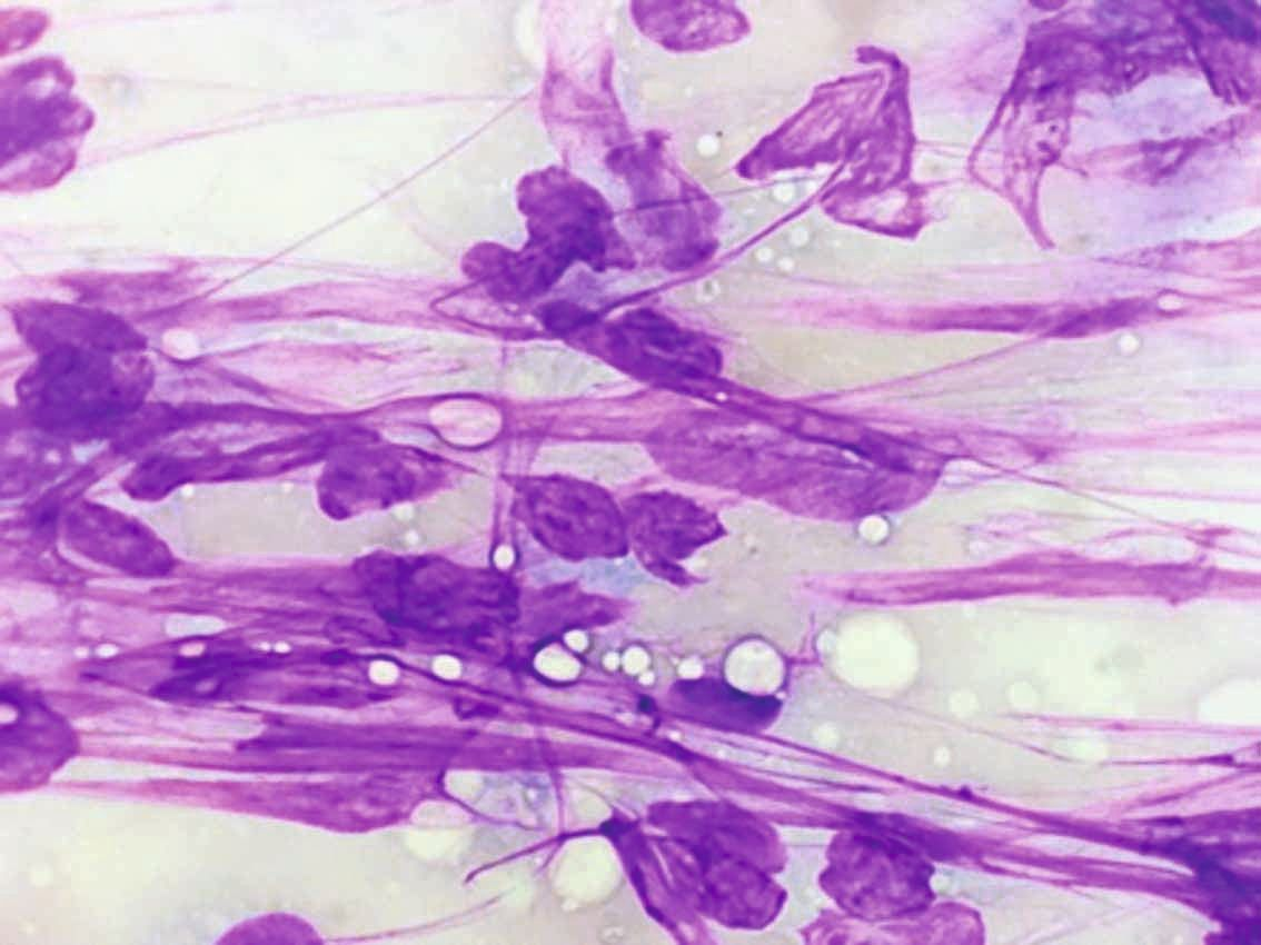
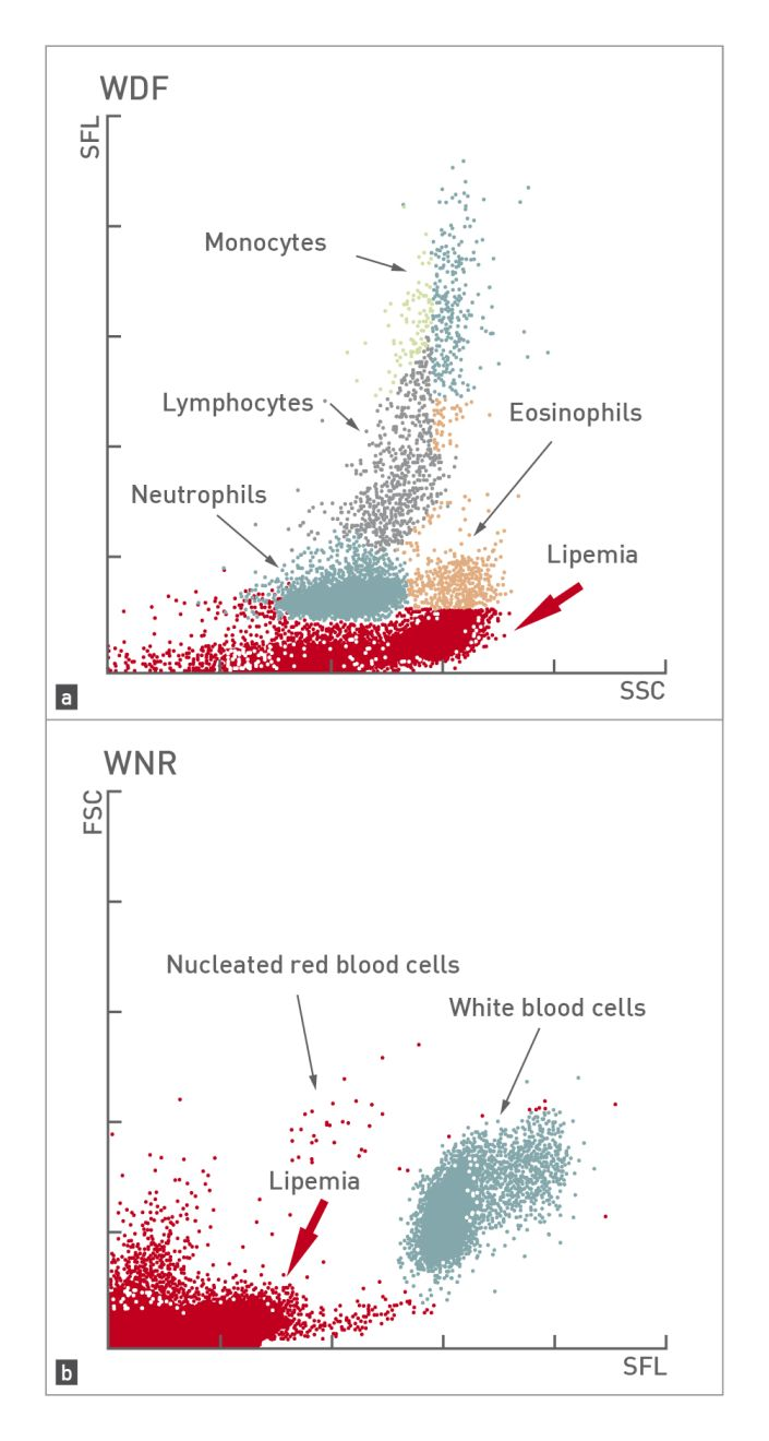
Lipemia (Figure 1) increases the turbidity of the sample and interferes with measurement of the hemoglobin concentration. In addition, depending on the analyzer operating system (impedance or laser beam), lipid microdroplets may lead to an erroneous increase in platelet count, mean corpuscular hemoglobin concentration (MCHC), and total leukocyte count [1],[2]. One of the most common causes of lipemia is a lack of fasting; therefore to reduce or eliminate this interference, samples must be obtained after the patient has been fasted for 12 hours.
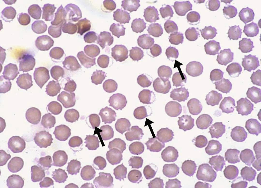
Laser beam hematology analyzers measure the erythrocyte count, hemoglobin concentration (both intracellular and free hemoglobin in plasma), and the mean corpuscular volume (MCV). If a sample is hemolyzed, the MCHC may be raised, while the erythrocyte count and hematocrit value are usually lower than normal [3]. MCHC is one of the most important erythrocyte parameters, since values above the reference range indicate errors or artifacts in plasma coloration (hemolysis, lipemia) or erythrocyte morphological changes (presence of spherocytes, eccentrocytes or Heinz bodies). For this reason, whenever a high MCHC is observed, the plasma sample and erythrocyte morphology must be evaluated (Figure 2).
Some laser beam analyzers can determine the cellular hemoglobin concentration mean (CHCM). This is calculated directly from the erythrocyte, not from the erythrocyte count and hemoglobin concentration, as is the case with MCHC. A discrepancy between the CHCM and MCHC values indicates interference in these parameters, with the most frequent cause being the presence of free hemoglobin in the sample from intravascular hemolysis and/or post-extraction [4],[5].
Should I consider the dog’s breed when interpreting hematological values?
Yes; ignorance of breed peculiarities can be problematic. For example, physiological macrothrombocytopenia (due to a mutation in the beta-1 tubulin gene) has been identified in the Cavalier King Charles Spaniel [6]. Analyzers detect these macroplatelets when calculating the plateletcrit (the volume occupied by platelets); this is important, as this measure is a better indicator of an adequate number of platelets than the actual platelet count [7]. If the mutation is suspected, definitive confirmation requires molecular testing.
There are many other examples. Some oriental breeds (e.g., the Akita Inu, the Shar-pei and the Shiba Inu) may present with physiological microcytosis (erythrocytes that are smaller than normal) and a reticulocyte hemoglobin value lower than the reference range for other breeds [8]. Poodles normally have a higher average corpuscular volume of erythrocytes than other breeds. Greyhounds, like other sighthounds, usually have a higher physiological hematocrit, erythrocyte count, and hemoglobin concentration, and a lower platelet count and mild leukopenia compared to other breeds [9].
Therefore when interpreting the results of an animal with respect to normal reference ranges, the species, age, and breed must always be taken into account. It is important to know the variations described in the dog breeds that are usually seen in the clinic.
Can I use heparin or citrate tubes for hematology?
Hematological analysis in the dog requires the use of whole blood anticoagulated with EDTA, but other anticoagulants, such as heparin or citrate, are also used in veterinary medicine, and incorrect anticoagulant use can lead to inaccurate results. Heparinized whole blood should not be used for cell counts since platelets and leukocytes frequently aggregate in such samples. Heparin anticoagulant is employed to obtain plasma for biochemical tests, whilst citrated whole blood is mainly used for coagulation assays and to obtain plasma or perform viscoelastic tests.
However, the use of citrated blood samples for cell counting has been described when EDTA samples have aggregation of platelets and/or leukocytes. When using citrate tubes (which contain 3.2% liquid sodium citrate) in this situation, the ratio of citrate-to-blood must be exactly 1:9. Because of this, citrate blood samples always have a slight hemodilution that must be taken into account in the values provided by the hematological analyzer [10].
EDTA is the anticoagulant of choice for hematological assays. In human medicine, it is advisable that the sample taken into the tube is within 10% of the volume recommended by the manufacturer. Under- or over-filling will affect the accuracy of hematology results. Excess EDTA will produce crenation of the erythrocytes and a decrease in their volume, which gives rise to a false reduction in microhematocrit and MCV [10].
Josep Pastor
How important is good quality control for a clinic’s hematology analyzer?
It is essential to have quality control programs available and to participate in them whenever working with an in-house hematology analyzer. The American Society of Veterinary Clinical Pathology (ASVCP) has published various guidelines with the aim of improving quality control in veterinary hematology [11],[12],[13],[14] and implementing improvements to prevent pre-analytical, analytical and post-analytical errors. Internal quality control programs must be followed using control samples, but in addition, participation in external, regional and/or international quality control programs is also necessary.
How do manual hematocrit values compare to those from an analyzer?
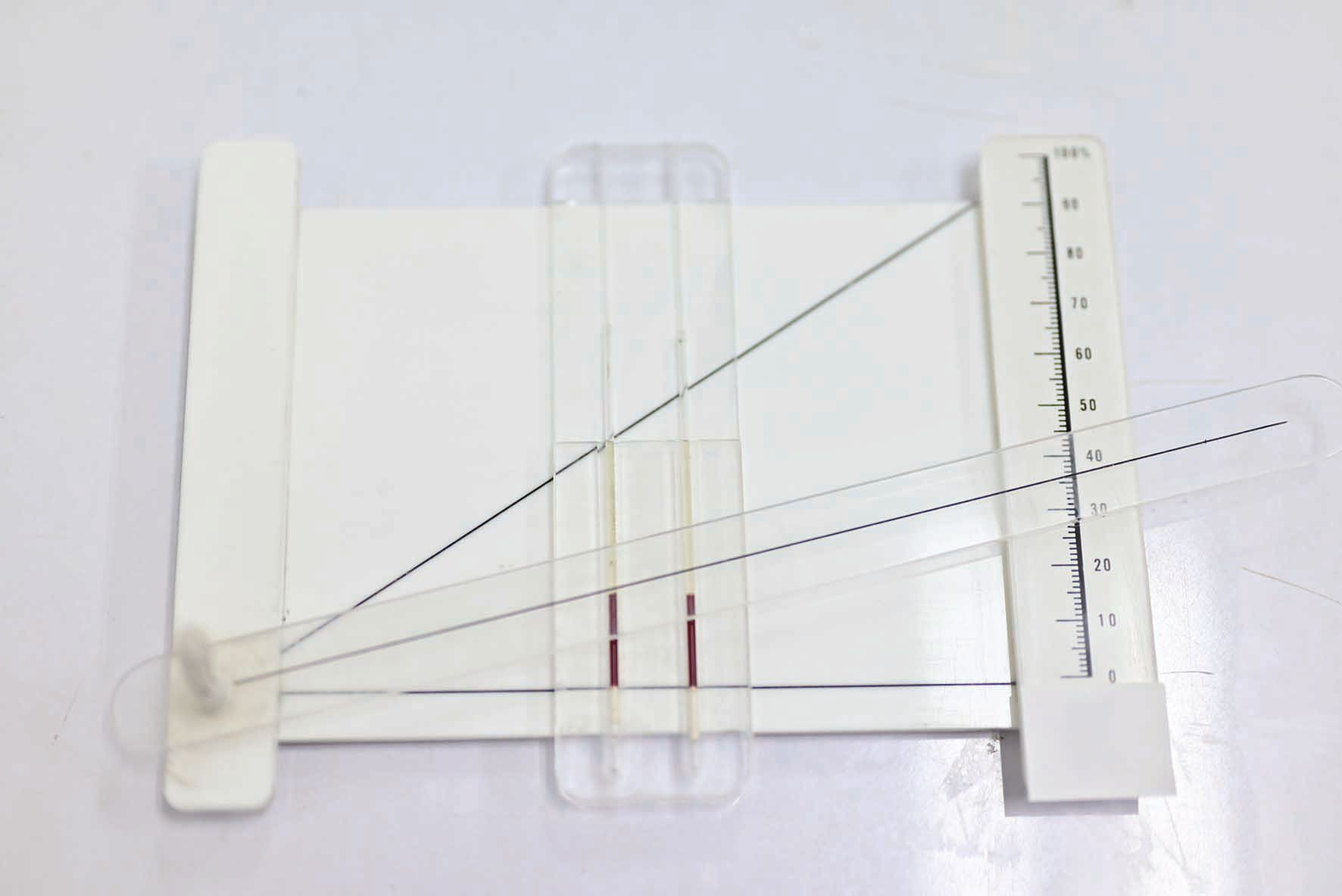
Automated hematology analyzers calculate the hematocrit value from the red blood cell count and MCV. However, a manual microhematocrit is the most accurate and reproducible method, done by centrifuging a sample at high speed and reading the capillary tube against a scale (Figure 3). Nevertheless, a recent study with veterinary students and veterinarians found that 25% of the students made errors related to inadequate mixing of blood prior to filling the tube, 23% read the results incorrectly, and 91% did not fill the microhematocrit tubes according to WHO recommendations [15],[16].
To reduce the number of samples requiring manual microhematocrit determination, it is advisable to study the relationship between hemoglobin concentration and hematocrit value. In general, the rule of 3 applies; the hematocrit value should be around 3 times the hemoglobin concentration. If the hematocrit value is outside this range, it is recommended that a manual microhematocrit should be performed and the result incorporated alongside the analyzer values for MCV and MCHC in the hematological analysis [10].
It is also worth mentioning that hypernatremia can alter the hematocrit value obtained with an automated analyzer. The erythrocytes of an animal with hypernatremia can swell when mixed with the analyzer diluent, falsely increasing the MCV and consequently the hematocrit [17].
Do hematology analyzers reliably identify left shift or toxic changes?
The ASVCP has established recommendations for microscopic review of blood smears and confirmation of automated differential leukocyte counts [14]. The opinions of veterinary experts regarding the evaluation of blood films or smears vary: some recommend all samples should include checking of the blood film, while others will advise that a review of the films and/or manual differentiation of leukocytes is carried out if certain specific criteria apply. The suitability of the different approaches depends on the patient population and context (i.e., sick patients, pre-anesthetic or geriatric examinations), the available hematology analyzer, and the knowledge base of the laboratory staff.
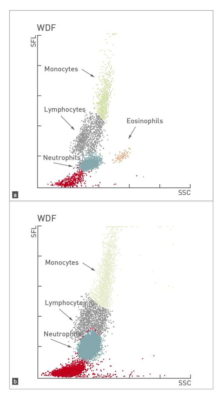
Laser beam hematology analyzers, in addition to producing numerical results, provide graphical presentations of the results (cytograms). Various veterinary studies have shown that cytograms are very useful as a deciding factor for reviewing blood smears [18],[19]. Taking this information into account, it is reasonable to recommend a study of the blood smear where;
- an abnormal cytogram is obtained;
- the analyzer flags a possible failure or error in the classification of leukocytes;
- the leukocyte count is outside the reference range.
Automatic laser beam analyzers can suggest the presence of toxic changes or a left shift (Figure 4); however, there is very little information on the sensitivity and specificity of this finding, so it is important to always review a blood smear for an animal that is sick or has leukocytosis, or when the analyzer issues an alert (Figure 5).
Can autoagglutination or parasites interfere with hematology analyzers?
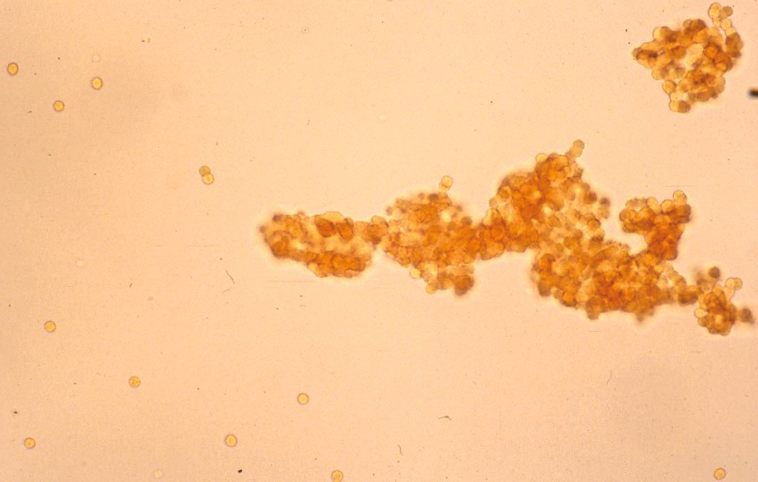
Macroscopic or microscopic autoagglutination (Figure 6) in blood samples is suggestive of an immune-mediated process; if found, the erythrocytes should be washed with physiological solution and the test repeated [20]. If the autoagglutination is genuine, aggregates can be observed in the analyzer cytogram, with the aggregates being counted as an individual cells, giving rise to an artificially low erythrocyte count [2].
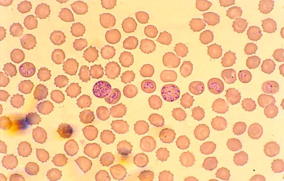
It has been shown that the detection of blood parasites such as Babesia spp. (Figure 7) causes changes in erythrocyte cytograms and may lead to an erroneous increase in the reticulocyte count. This is because laser beam analyzers employ fluorescent polymethine stain, and parasites present within the erythrocytes can also be stained [21],[22]. However, this is rare, and the sensitivity of these analyzers in detecting blood parasites is unknown, so it is currently recommended to evaluate the blood smear if there is anything suspicious, or to do so routinely in animals from areas where babesiosis or other vector-borne diseases are endemic [23],[24].
Should thrombocytopenia on a hematology analyzer be confirmed with a blood smear?
Whenever a decreased platelet count is found, examination of a blood smear is indicated – partly as artifacts due to transport of the sample or problems during blood sampling are common in veterinary medicine. The platelet count can be estimated on a blood smear by multiplying the average number of platelets per field at 1000× (oil immersion) × 15000/µL. However, a study showed that the variability of estimated platelet counts in dogs using this method is very high and depends on the operator and the smear area evaluated [25].
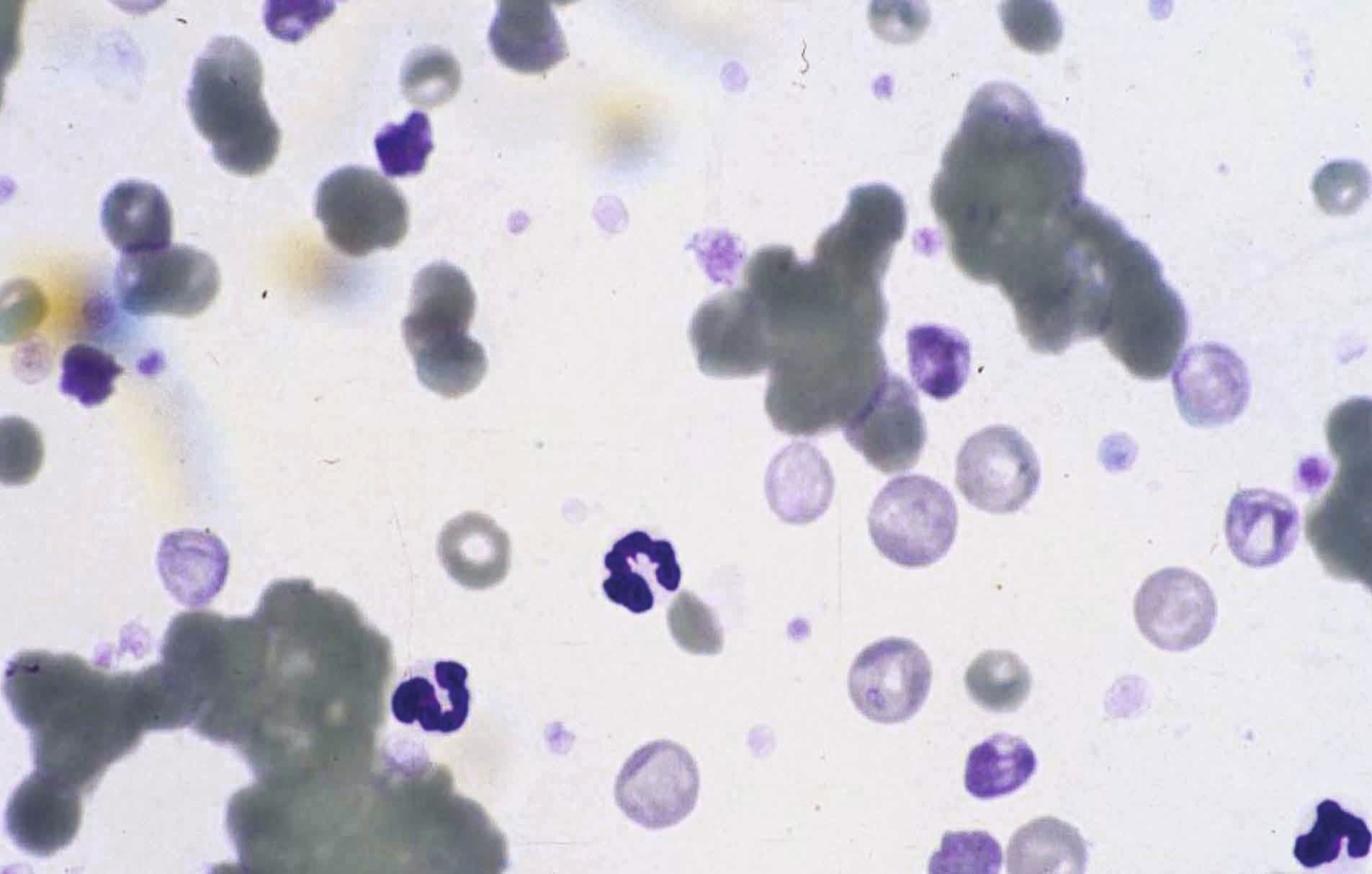
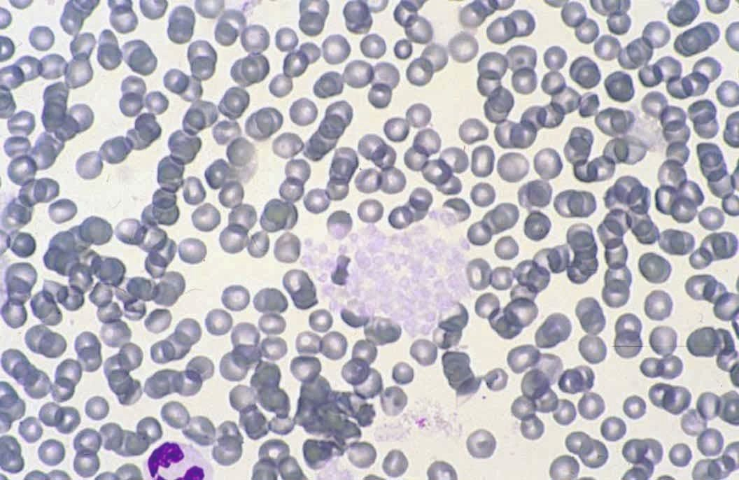
Invalidating platelet counts in all samples with platelet aggregates is frustrating and possibly unnecessary for most hematological samples. According to clinical recommendations, if platelet counts are within the reference ranges, platelet aggregates are clinically insignificant. However, whenever an automated count indicates thrombocytopenia, a blood smear should be reviewed to evaluate platelet aggregates (Figure 8) [26]. Newer laser hematology analyzers have indicators for platelet aggregates, and depending on the number and size of the aggregates, the clinician may be able to detect their presence in the cytograms and histograms provided by the analyzer. This can help reduce the need to examine blood smears in clearly defined cases.
It is worth noting that a recent article evaluated the determination of immature platelet fraction (IPF) in dogs using a commercial analyzer. These immature cells are an indicator of bone marrow regeneration of platelets when there is greater demand. The study reported that the values provided by the analyzer were slightly overestimated in the presence of platelet aggregates [27].
Conclusion
Veterinary hematology has advanced significantly in recent years, especially due to the incorporation of more technologically advanced analyzers. However, the clinician should be aware that there are many factors which can lead to erroneous or misleading results; good sampling technique and careful processing are mandatory for reliable diagnosis, along with regular and appropriate quality control procedures for the in-house analyzers. Manual examination of blood smears is still necessary on many occasions for better diagnosis of our pets!

Josep Pastor
DVM, PhD, Dip. ECVCP, Faculty of Veterinary Medicine, Barcelona Autonomous University, Barcelona, SpainSpain
Josep Pastor obtained both his bachelor’s degree and doctorate in veterinary medicine from the Barcelona Autonomous University (UAB) and has been a professor of Medical Pathology at the UAB Faculty of Veterinary Medicine since 1991. He is also currently co-director of the Hematology Unit in the University’s Department of Animal Medicine and Surgery. He attained diplomate status from the European College of Veterinary Clinical Pathology in 2002 and has authored several veterinary textbooks, as well as numerous articles for both national and international journals. His main areas of interest are hematology, clinical pathology and small animal oncology.
References
- Zandecki M, Genevieve F, Gerard J, et al. Spurious counts and spurious results on haematology analysers: a review. Part I: platelets. Int. J. Lab. Hematol. 2007;29(1):4-20.
- Zandecki M, Genevieve F, Gerard J, et al. Spurious counts and spurious results on haematology analysers: a review. Part II: white blood cells, red blood cells, haemoglobin, red cell indices and reticulocytes. Int. J. Lab. Hematol. 2007;29(1):21-41.
- Berda-Haddad Y, Faure C, Boubaya M, et al. Increased mean corpuscular haemoglobin concentration: artefact or pathological condition? Int. J. Lab. Hematol. 2017;39(1):32-41.
- Stockham SL, Scott MA. Fundamentals of Veterinary Clinical Pathology, 2nd ed. Ames, Iowa: Blackwell, 2008. p120-122
- Stokol T, Erb HN. A comparison of platelet parameters in EDTA‐ and citrate‐anticoagulated blood in dogs. Vet. Clin. Pathol. 2007;36:148-154.
- Davis B, Toivio-Kinnucan M, Schuller S, et al. Mutation in beta1-tubulin correlates with macrothrombocytopenia in Cavalier King Charles Spaniels. J. Vet. Intern. Med. 2008;22(3):540-545.
- Tvedten HW, Lilliehöök IE, Oberg J, et al. Validation of Advia plateletcrit for assessing platelet mass in dogs, including Cavalier King Charles spaniels. Vet. Clin. Pathol. 2012;41(3):336-343.
- Schaefer DM, Stokol T. The utility of reticulocyte indices in distinguishing iron deficiency anemia from anemia of inflammatory disease, portosystemic shunting, and breed-associated microcytosis in dogs. Vet. Clin. Pathol. 2015;44(1):109-119.
- Zaldívar-López S, Marín LM, Iazbik MC, et al. Clinical pathology of Greyhounds and other sighthounds. Vet. Clin. Pathol. 2011;40(4):414-425.
- Harvey JW. Veterinary Hematology: A Diagnostic Guide and Color Atlas. St. Louis, MO: Elsevier/Saunders, 2012. p17-19
- Arnold JE, Camus MS, Freeman KP, et al. ASVCP Guidelines: Principles of Quality Assurance and Standards for Veterinary Clinical Pathology (version 3.0). Vet. Clin. Pathol. 2019;48(4):542-618.
- Flatland B, Freeman KP, Friedrichs KR, et al. ASVCP quality assurance guidelines: control of general analytical factors in veterinary laboratories. Vet. Clin. Pathol. 2010;39(3):264-277.
- Camus MS, Flatland B, Freeman KP, et al. ASVCP quality assurance guidelines: external quality assessment and comparative testing for reference and in-clinic laboratories. Vet. Clin. Pathol. 2015;44(4):477-492.
- Vap LM, Harr KE, Arnold JE, et al; ASVCP quality assurance guidelines: control of preanalytical and analytical factors for hematology for mammalian and nonmammalian species, hemostasis, and crossmatching in veterinary laboratories. Vet. Clin. Pathol. 2012;41(1):8-17.
- Breheny CR, Brown A, Handel I, et al. Inter- and intra-operator variability in the analysis of packed cell volume. J. Small Anim. Pract. 2017;58(1):29-34.
- World Health Organization Diagnostic Imaging and Laboratory Technology. Recommended method for the determination of packed cell volume by centrifugation. World Health Organization, Geneva, 2000
- Boisvert AM, Tvedten HW, Scott MA. Artifactual effects of hypernatremia and hyponatremia on red cell analytes measured by the Bayer H*1 analyzer. Vet. Clin. Pathol. 1999;28(3):91-96.
- Lilliehook I, Tvedten H. Validation of the Sysmex XT‐2000iV hematology system for dogs, cats, and horses. II. Differential leukocyte counts. Vet. Clin. Pathol. 2009;38:175-182.
- Stirn M, Moritz A, Bauer N. Rate of manual leukocyte differentials in dog, cat and horse blood samples using ADVIA 120 cytograms. BMC Vet. Res. 2014;10:125.
- Garden OA, Kidd L, Mexas AM, et al. ACVIM consensus statement on the diagnosis of immune-mediated hemolytic anemia in dogs and cats. J. Vet. Intern. Med. 2019;33(2):313-334.
- Piane L, Théron ML, Aumann M, et al. Spurious reticulocyte profiles in a dog with babesiosis. Vet. Clin. Pathol. 2016;45(4):594-597. Doi: 10.1111/vcp.12395.
- Piane L, Young KM, Giraud L, et al. Spurious reticulocyte profiles in dogs with large form babesiosis: a retrospective study. Vet. Clin. Pathol. 2016;45(4):598-603. Doi: 10.1111/vcp.12396.
- Bauer N, Nakagawa J, Dunker C, et al. Evaluation of the automated hematology analyzer Sysmex XT-2000iV™ compared to the ADVIA® 2120 for its use in dogs, cats, and horses: Part I – Precision, linearity, and accuracy of complete blood cell count. J. Vet. Diagn. Invest. 2011;23(6):1168-1180.
- Bauer N, Nakagawa J, Dunker C, et al. Evaluation of the automated hematology analyzer Sysmex XT-2000iV™ compared to the ADVIA® 2120 for its use in dogs, cats, and horses. Part II – Accuracy of leukocyte differential and reticulocyte count, impact of anticoagulant and sample aging. J. Vet. Diagn. Invest. 2012;24(1):74-89.
- Paltrinieri S, Paciletti V, Zambarbieri J. Analytical variability of estimated platelet counts on canine blood smears. Vet. Clin. Pathol. 2018;47:197-204.
- Tvedten HW, Backlund K, Lilliehook IE. Reducing error in feline platelet enumeration by addition of Iloprost to blood specimens: comparison to prostaglandin E1 and EDTA. Vet. Clin. Pathol. 2015;44:179-187.
- Jornet-Rius O, Mesalles-Naranjo M, Pastor J. Performance of the Sysmex XN-V hematology analyzer in determining the immature platelet fraction in dogs: A preliminary study and reference values. Vet. Clin. Pathol. 2023;52(3):433-442.