Introduction
Round cell tumors are diverse neoplasms commonly encountered in veterinary medicine, particularly in dogs, with the category including transmissible venereal tumors (TVT), lymphomas, mast cell tumors, plasma cell tumors, and histiocytomas. When viewed under a microscope, these tumors are characterized by a round appearance of the cells, but each has distinct cytologic appearance and clinical features. This article offers a brief guide for the first opinion clinician in what to look for when doing initial investigations, and also what features can be misleading and possibly lead to an erroneous diagnosis.
Transmissible venereal tumor (TVT)
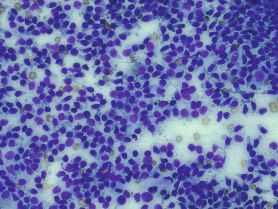
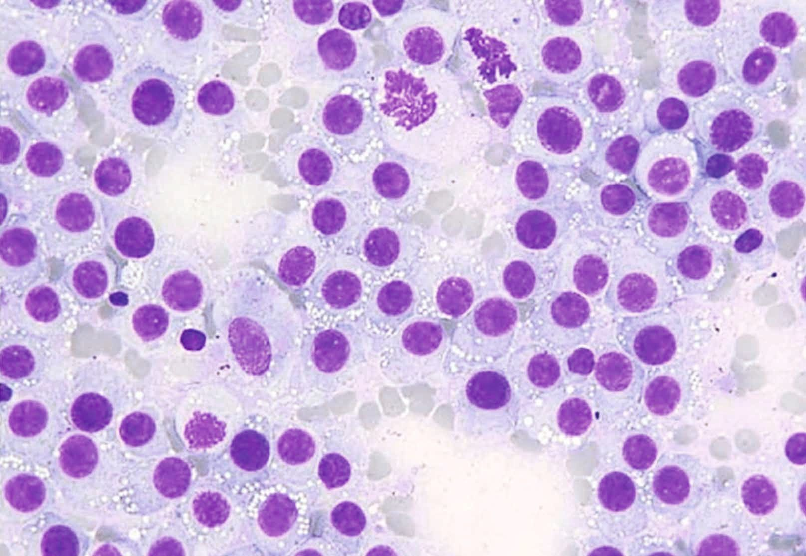
TVT is a unique type of contagious cancer that affects dogs, transmitted through direct contact, usually during mating. It predominantly affects the external genitalia but can also appear in extragenital locations. TVT cells are distinctive with their cytoplasmic vacuoles; these are discrete and punctate, and are frequently described as a “string of pearls” (Figure 1).
Lymphoma
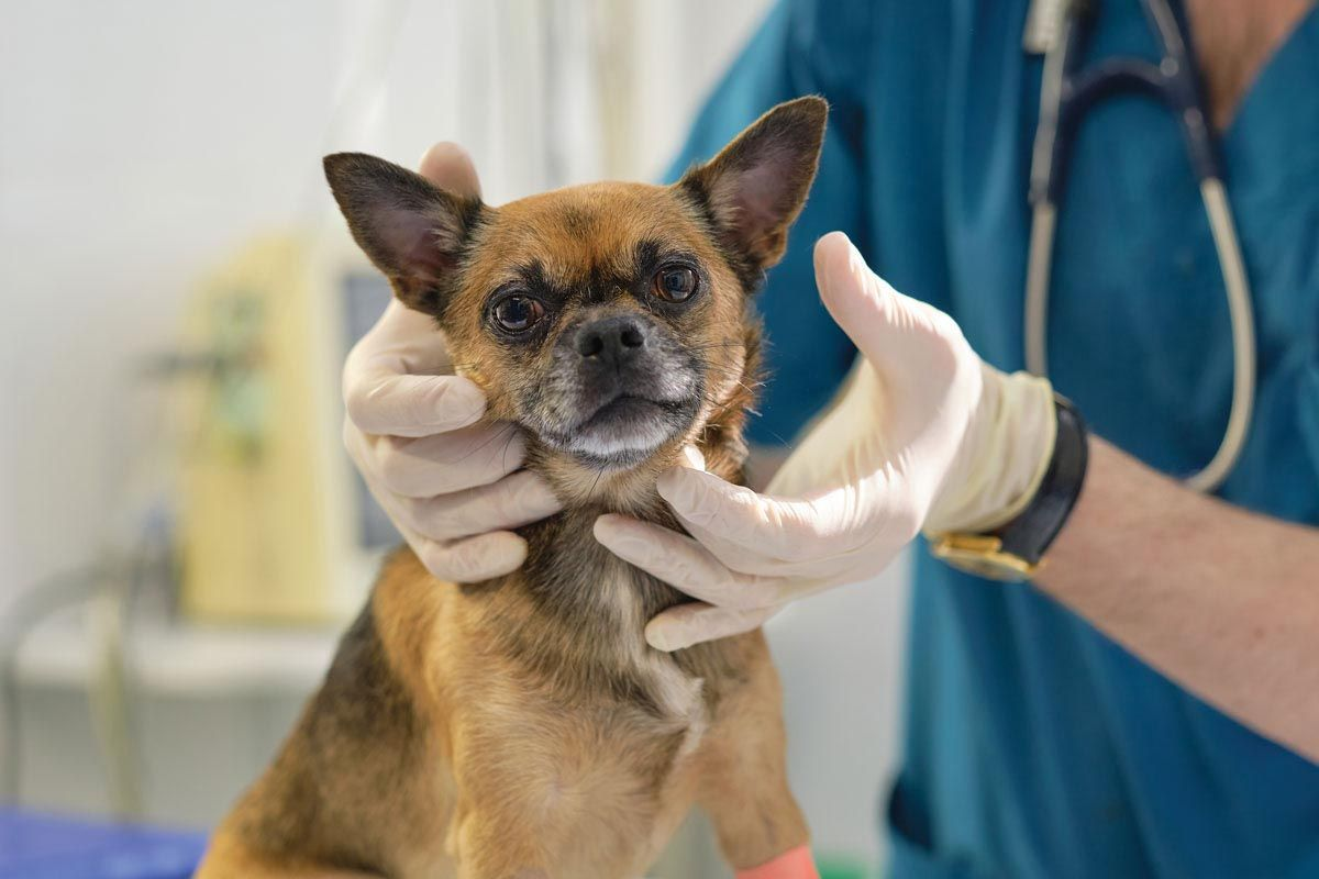
Lymphoma in dogs is a common presentation, and a preliminary diagnosis can be made on clinical examination (Figure 2), but it is recommended to make identification definitive, as this will help in advising the owner on treatment and prognosis. In cytology reports, lymphoma is often diagnosed as “intermediate cell lymphoma,” “intermediate to large cell lymphoma,” or “large cell lymphoma”. These terms may lead to a misconception that “large cell lymphoma” is a distinct disease type, but in fact there are more than 30 types of lymphoma based on WHO classification [1]. Many of them could be diagnosed as “large cell lymphoma”, but the presentation and exact immunophenotype can vary widely, from indolent to aggressive forms, and hence the prognosis will also vary correspondingly. Discussion here will center on a few lymphomas that have distinct cytologic features which may be recognized on cytology without ancillary tests, as an understanding of these features may provide a rough idea of possible diagnosis and prognosis; however, the complete classification and a definitive diagnosis of canine lymphoma can only be achieved by combining cytology, histopathology, immunohistochemistry, and advanced molecular testing (e.g., flow cytometry).
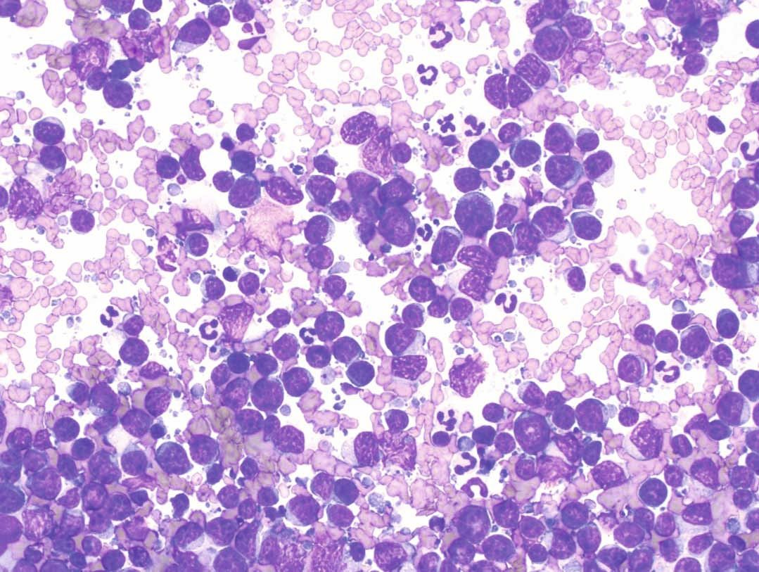
- The large or intermediate to large cell lymphoma: This type of lymphoma primarily comprises lymphocytes that are intermediate to large (more than 2-3 times the size of red blood cells). This is the most common diagnosis of lymphoma seen on a cytology report. Typically, large neoplastic lymphocytes have a high nuclear-to-cytoplasmic ratio (N:C ratio) and small to scant amounts of basophilic cytoplasm (Figure 3). Based on the clinical presentation and the immunophenotype, the lymphoma could be a “Diffuse Large B Cell Lymphoma (DLBCL)” or “Peripheral T Cell Lymphoma Not Otherwise Specified (PTCL-NOS)”.
- Diffuse Large B Cell Lymphoma (DLBCL): DLBCL is the most common canine lymphoma, and it generally affects middle-aged to older dogs, with a median age of onset around 7-9 years. Certain breeds, including Golden Retrievers, Labrador Retrievers, Bernese Mountain Dogs, and German Shepherds, are at higher risk. Dogs with lymphoma typically present with generalized lymphadenopathy. The median survival time is approximately 300-500 days [2].
- Peripheral T-Cell Lymphoma (PTCL-NOS): PTCL-NOS accounts for roughly 15% of all canine lymphoma cases. This type tends to present in middle-aged Boxers and is slightly more common in male dogs. The disease often involves the mediastinal lymph nodes and can be associated with hypercalcemia. Unfortunately, PTCL-NOS is associated with a poor prognosis, with a median survival time of only about 158 days [2].
Given the cytologic similarity and prognostic difference between DLBCL and PTCL-NOS, flow cytometry or histopathology with immunohistochemistry is strongly recommended to confirm the diagnosis.
- T-Zone Lymphoma (TZL): TZL is a small to intermediate cell lymphoma that has a better prognosis than other types. It often presents with a “hand mirror” or “tadpole” appearance cytologically (Figure 4). However, flow cytometry is necessary for definitive diagnosis, as TZL cells will typically be positive for CD3, CD5, CD21, and MHC II cell markers [3]. This type of lymphoma frequently involves the blood, leading to lymphocytosis. Golden Retrievers, Maltese Terriers and Shih Tzus are breeds commonly affected. The indolent nature of TZL means a better median survival time, which can be as long as 637-760 days [3].
- Large Granular Lymphocyte (LGL) Lymphoma: LGL is characterized by cytotoxic T cells or natural killer (NK) cells that often have pink to magenta cytoplasmic granules containing enzymes such as perforin and granzymes. In dogs this type of lymphoma is primarily found in the liver and spleen, but it can also be present in the gastrointestinal tract or as a disseminated disease. Diagnosis is typically confirmed through cytology, because histopathology, which does not show cytoplasmic granules well, will often indicate a low-grade lymphoma. The aggressive nature of the disease is indicated by a median survival time of only 28 days, even though around 75% of dogs may initially respond to chemotherapy. A positive prognostic factor includes finding an intermediate lymphocyte size (versus large size) on cytology, which correlates with a median survival time of more than 100 days in some patients [4].
- Marginal Zone Lymphoma (MZL): MZL is best diagnosed through histopathology, although the cytologic feature of a single prominent nucleolus can be indicative. This type of lymphoma can have a variable prognosis depending on the affected organ. In the spleen, it can have a good long-term prognosis with splenectomy alone, whereas lymph node involvement can be as aggressive as DLBCL [2].
Mast cell tumor (MCT)
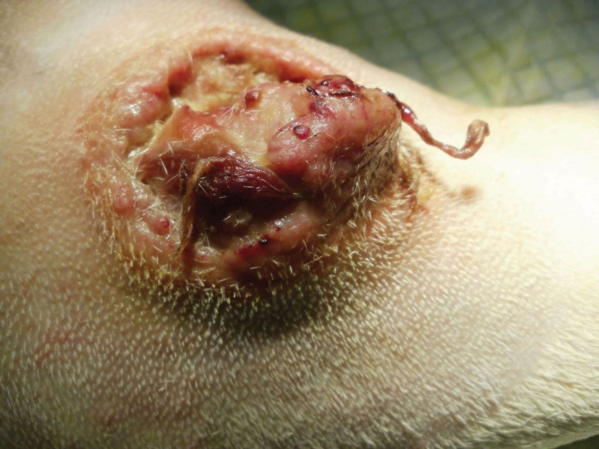
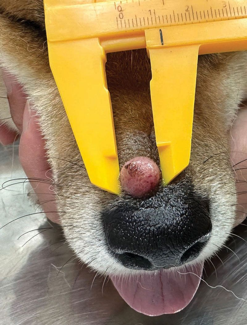
These are the most common skin tumors in dogs, representing 16.8% of all cutaneous neoplasms [5]. The mean age for MCT presentation is around nine years, and they can occur in any breed, but some are predisposed; these include Boxers, Pugs, Labrador Retrievers, Golden Retrievers, and Weimaraners. The tumors are often solitary but they can be multiple, and are typically found on the trunk (50%), limbs (40%), and head and neck (10%). The clinical appearance can range from erythematous alopecic masses or plaques to raised nodules (Figure 5) [5].
Cytological features
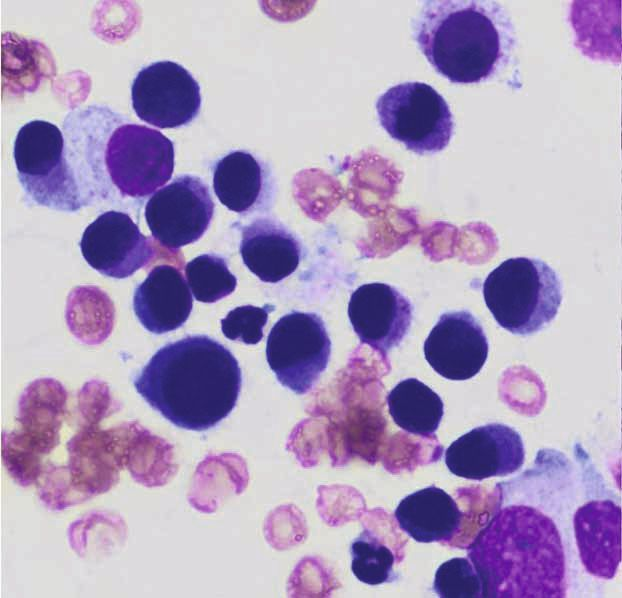
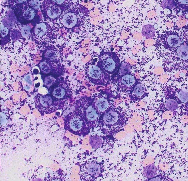
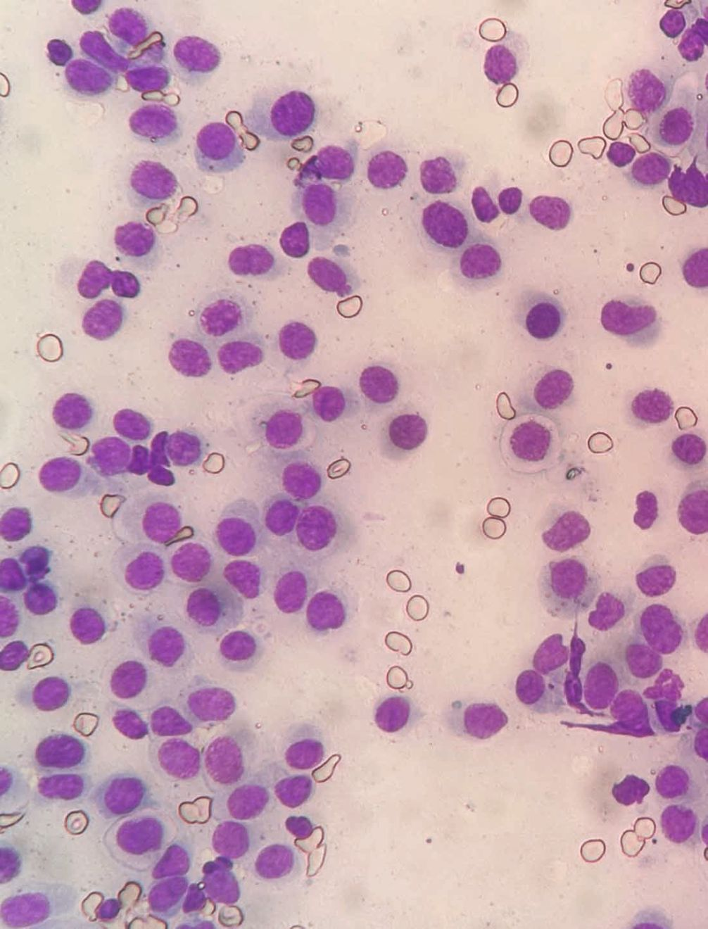
MCT are characterized cytologically by centrally located round nuclei with purple intracellular granules that vary in size and number. Note that the granules may not be visible if using an in-clinic aqueous Romanowsky stain (e.g., Diff-QuikTM stain) (Figure 6). Other components commonly associated with MCTs include collagen fibers, eosinophils, and fibroblasts.
Cytologic grading
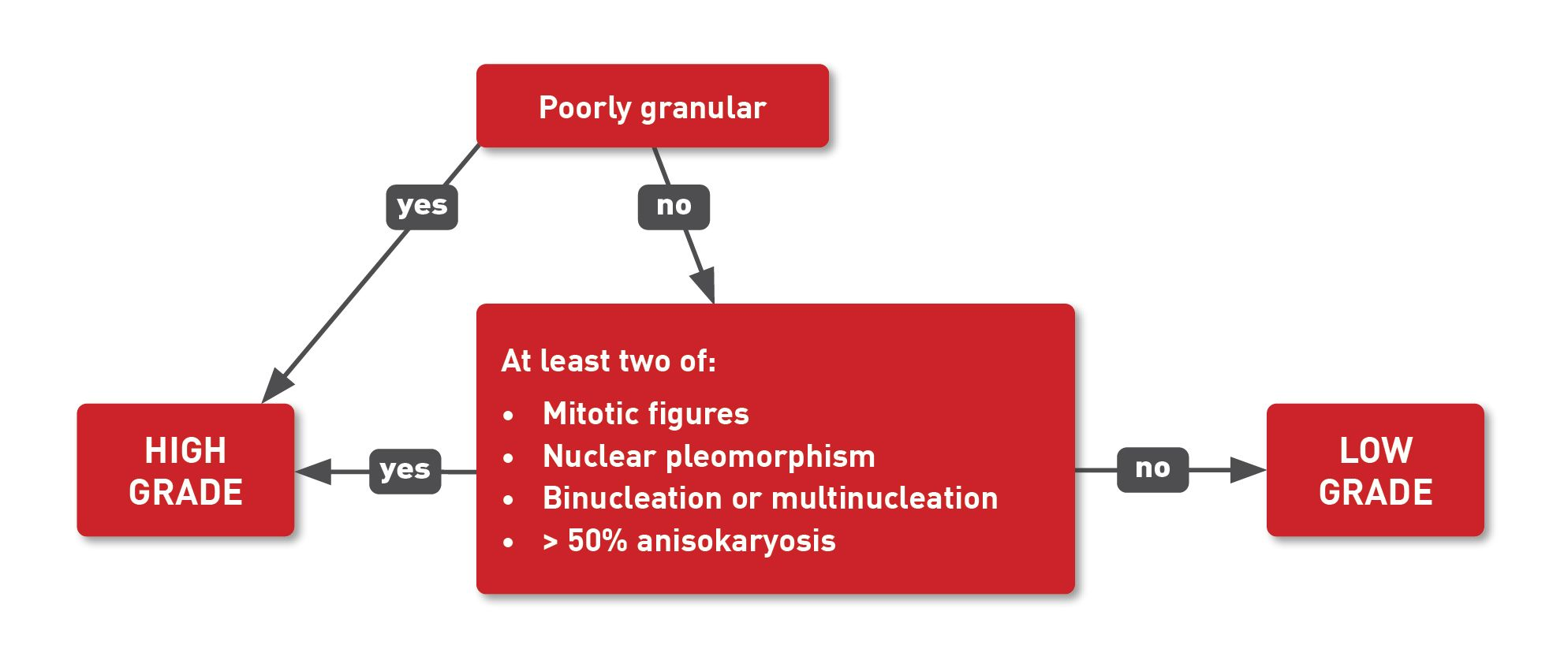
Tumor grading assesses how normal or abnormal the cancer cells look under a microscope, and cytologic grading is a crucial aspect of diagnosing MCT. Generally, the more abnormal the cells appear, the more aggressive the cancer is, and the faster it is likely to grow and spread. The cytologic grading for canine cutaneous MCT involves examining specific criteria based on morphologic characteristics of neoplastic cells, including karyomegaly, multinucleation, nuclear pleomorphism, and mitotic figures. A recent paper has described using cytological identification of granulation alongside a set of criteria to determine the grading of a MCT [6]. Essentially, poor granulation on cytologic evaluation indicates high grade, but if the cells are well-granulated, fulfilling at least two of the following criteria would also indicate a high-grade MCT (Box 1):
- Presence of mitotic figures
- Nuclear pleomorphism (variation in nuclear size and shape)
- Binucleation or multinucleated cells
- Anisokaryosis, which refers to a significant (> 50%) variation in nuclear size
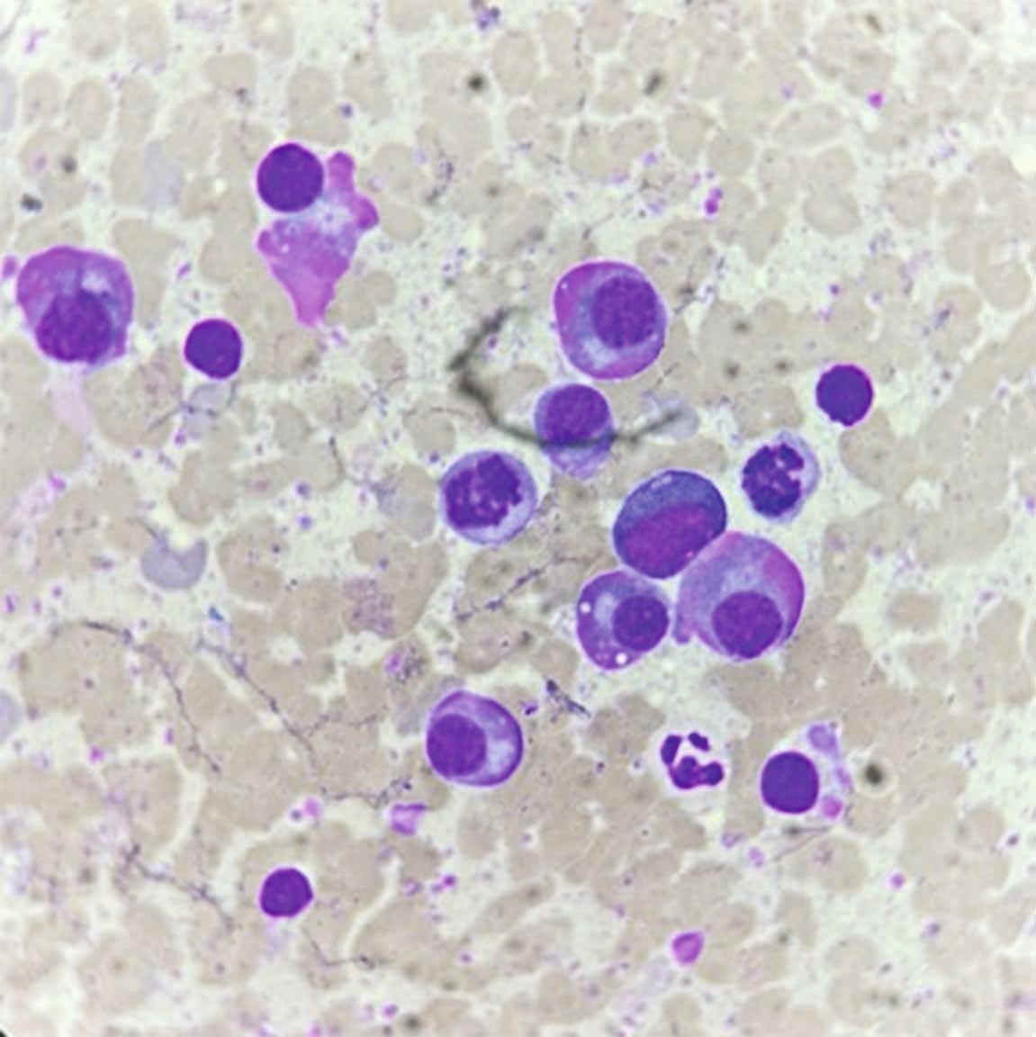
A recent study suggested a new development for cytologic grading [7]. It reported that examination of slides with a Romanowsky stain could allow inclusion of additional factors for grading, such as fibroblasts and collagen fibrils. A moderate to intense concentration of these elements suggests a more benign character associated with increased survival and a lower histopathologic grade.
Candice P. Chu
Plasma cell tumor
Also known as plasmacytoma, plasma cell tumors in dogs are regarded as benign neoplasms that can appear “ugly” on cytology. These tumors often occur in middle-aged to older dogs, with common sites including the limbs and areas of the head such as the lip and ear margins. Cytologically, they have a characteristic perinuclear clearing known as the Golgi zone and may present bi- and multi-nucleation. Their nuclei often have clumped chromatin, often described as “clock-face” or “soccer ball” in appearance. Occasionally, plasma cells may have a pink fringe known as “flame cells” (Figure 7).
Histiocytoma
Histiocytomas are benign skin tumors most commonly found in younger dogs under two years old. They appear as solitary, firm, dome-shaped masses primarily located on the head and limbs, and are often ulcerated. Under the microscope these tumors show a “fried egg” appearance, with pale blue to colorless cytoplasm and few mitotic figures (Figure 8). Histiocytomas often regress spontaneously without treatment.
Conclusion
Round cell tumors in dogs comprise a variety of neoplasms that are relatively common in veterinary practice. Each type has unique characteristics that require specific diagnostic and treatment approaches. Understanding the cytological features of these tumors is critical for veterinarians to provide accurate diagnoses, prognoses, and therapeutic strategies to manage these conditions effectively in canine patients.

Candice P. Chu
DVM, PhD, Dip. ACVP (Clinical Pathology), Assistant Professor, Texas A&M University, TX, USA
United States
Dr. Chu is a board-certified veterinary clinical pathologist and a tenure-track assistant professor at Texas A&M University. She earned her DVM at the National Taiwan University in Taipei City, Taiwan, and her PhD in veterinary pathobiology at Texas A&M University, where she also completed residency training. Her research interests include gene and microRNA expression of chronic kidney disease, focusing on urinary microRNAs as biomarkers of kidney diseases in dogs and cats, and artificial intelligence (AI) in digital cytology and clinical pathology. As an innovative educator, she founded the Instagram page Vet.Clin.Path.Professor and teaches cytology to veterinary students and veterinarians worldwide.
References
- Valli VE, San Myint M, Barthel A, et al. Classification of canine malignant lymphomas according to the World Health Organization criteria. Vet. Pathol. 2011;48:198-211.
- Avery AC. The genetic and molecular basis for canine models of human leukemia and lymphoma. Front. Oncol. 2020;10:23.
- Seelig DM, Avery P, Webb T, et al. Canine T-zone lymphoma: unique immunophenotypic features, outcome, and population characteristics. J. Vet. Intern. Med. 2014;28:878-886.
- Yale AD, Crawford AL, Gramer I, et al. Large granular lymphocyte lymphoma in 65 dogs (2005-2023). Vet. Comp. Oncol. 2023 (pub on line https://doi.org/10.1111/vco.12959)
- London CA, Thamm DH. Mast cell tumor. In: Vali DM, Thamm DH, Liptak JM (eds.) Withrow and MacEwens Small Animal Clinical Oncology E-Book; Elsevier Health Sciences; 2019;335-355.
- Camus M, Priest H, Koehler J, et al. Cytologic criteria for mast cell tumor grading in dogs with evaluation of clinical outcome. Vet. Pathol. 2016;53:1117-1123.
- Paes PR, Horta RS, Luza LC, et al. Inclusion of fibroblasts and collagen fibrils in the cytologic grading of canine cutaneous mast cell tumors. Vet. Clin. Pathol. 2022;51:339-348.