Vitamin D synthesis and metabolism
In many species, the biosynthesis of vitamin D begins with exposure to UV light, whereby 7-dehydrocholesterol is transformed to previtamin D3. Factors that affect synthesis of vitamin D3 include quantity and quality of the UV light, the animal’s coat type, and skin pigmentation. Dogs differ from humans (and many other species) in that they lack the ability to synthesize vitamin D3 in the skin, likely because of high activity of the enzyme 7-dehydrocholesterol-Δ7-reductase. For this reason, dogs require dietary supplementation with vitamin D to meet nutritional requirements. There are two dietary forms of vitamin D: cholecalciferol (vitamin D3 ), which typically comes from animal food sources, and ergocalciferol (vitamin D2 ), which typically comes from plant sources.Vitamin D is supplied in commercial dog foods in the form of various ingredients (e.g., organ meat or oily fish products) and supplemental cholecalciferol. Current AAFCO1 recommendations for minimum and maximum amounts of dietary vitamin D intake are 125 IU and 750 IU per 1000 kilocalories, respectively. While the cholecalciferol concentration in most commercially available diets has minimal impact on a dog’s serum 25(OH)D concentration, it can, if given in high enough quantities (up to 2700 IU/kg body weight) impact serum 25(OH)D concentrations [1]. Clinicians should be aware that this dose far exceeds the National Research Council (NRC) safe upper limit of 2.6 μg (i.e., 104 IU) per kg body weight (BW)0.75.
Once ingested, vitamin D is transported to the liver via the portal system and intestinal lymphatics ( Figure 1 ). This process requires digestive enzymes, chylomicrons, bile acids, vitamin D-binding protein (VDBP), and transcalciferin. In the liver, cholecalciferol is hydroxylated by 25-hydroxylase to form 25(OH)D (also known as calcidiol or calcifediol), which binds to VDBP in the circulation. With a half-life of approximately 2 to 3 weeks, 25(OH)D is thought to be the most reliable indicator of systemic vitamin D status.
25(OH)D is then hydroxylated (by 1α-hydroxylase) to form 1,25(OH)2 D (also known as calcitriol), the most active naturally occurring vitamin D metabolite; this affects many target cells via a vitamin D receptor (VDR)–mediated mechanism ( Figure 1 ). Calcitriol binds to the VDR much more readily (approximately 500 times) than vitamin D3 or 25(OH)D. This activation of 1,25(OH)2 D occurs predominantly in the kidneys but also in other tissues that express 1α-hydroxylase. In dogs, VDR expression has been identified in several tissues, especially the kidney, duodenum, skin, ileum, and spleen. Although the exact mechanism has not been completely elucidated, 1α-hydroxylase activity is tightly regulated by serum concentrations of calcium, parathormone (PTH), 1,25(OH)2 D, fibroblast growth factor 23 (FGF-23), and activity of the enzyme Klotho. Within cells, 1,25(OH)2 D can promote or suppress gene transcription and expression. Both 25(OH)D and 1,25(OH)2 D are inactivated via 24-hydroxylase to form 24,25(OH)2 D and 1,24,25-trihydroxyvitamin D respectively, and other metabolites (e.g., 25[OH]D-23,23 lactone) that are excreted in the urine and bile.
1 AAFCO – Association of American Feed Control Officials
Vitamin D roles
Classically, vitamin D is known for its influence on calcium-phosphorus homeostasis via the bone-parathyroid-kidney axis. However, vitamin D has multiple other effects throughout the body, as evidenced by the wide variety of cells that express the VDR. Actions induced by VDR activation in humans include differentiation of immune cells, reductions in inflammation and proteinuria, increased insulin secretion, and improvement of hematopoiesis.Measuring vitamin D metabolites
There are no universally accepted “normal” reference ranges for vitamin D metabolites. Part of the difficulty in interpreting laboratory results relates to the fact that multiple techniques are employed to measure the metabolites; these include liquid chromatographic methods, immunoassay techniques, chemiluminescence immunoassays, and radioimmunoassays. There can be significant inter-assay, intra-assay, and interlaboratory variance. In an effort to assist in the development of standard reference materials and to examine differences among assay performance the National Institute of Standards and Technology (NIST) and the National Institutes of Health (NIH) Office of Dietary Supplements (ODS) established a Vitamin D Metabolites Quality Assurance Program (VitDQAP). Comparability of vitamin D metabolite measurements has improved greatly over time via development of these quality control efforts; however, the studies were performed with human samples, and the effect of a canine or feline matrix on these variables and comparability of results is unknown.
Liquid chromatography assays are currently the most commonly used methods and remain the criterion-referenced standard (liquid chromatography with tandem mass spectrometric detection) for measurement. Wherever possible, it is recommended to use a laboratory that has received certification either from The Centers for Disease Control and Prevention (CDC) Vitamin D Standardization-Certification Program (VDSCP) and/ or the Vitamin D External Quality Assessment Scheme (DEQAS) to increase the likelihood of accurate results.
How much vitamin D is enough?
Defining 25(OH)D sufficiency, insufficiency, and deficiency is controversial. In humans, vitamin D deficiency is generally defined as < 20 ng/mL and sufficiency is generally >30 ng/mL. Optimal repletion is defined by some as > 50 or > 60 ng/mL to achieve the aforementioned pleiotropic effects on the VDR. Multiple variables (including signalment, disease, assay technique, and physiologic variation) affect the reference range and the therapeutic target range.Consensus on optimal, adequate, or deficient vitamin D status in healthy canine populations has not been determined. Wide ranges of 25(OH)D concentrations have been reported for healthy dogs, and there is no universally accepted “normal” range – and importantly, assay choice and technique differ among many of these studies. In one study of apparently healthy dogs, the circulating 25(OH)D concentrations varied markedly, from 9.5 to 249 ng/mL [ Vitamin D metabolite status in various diseases]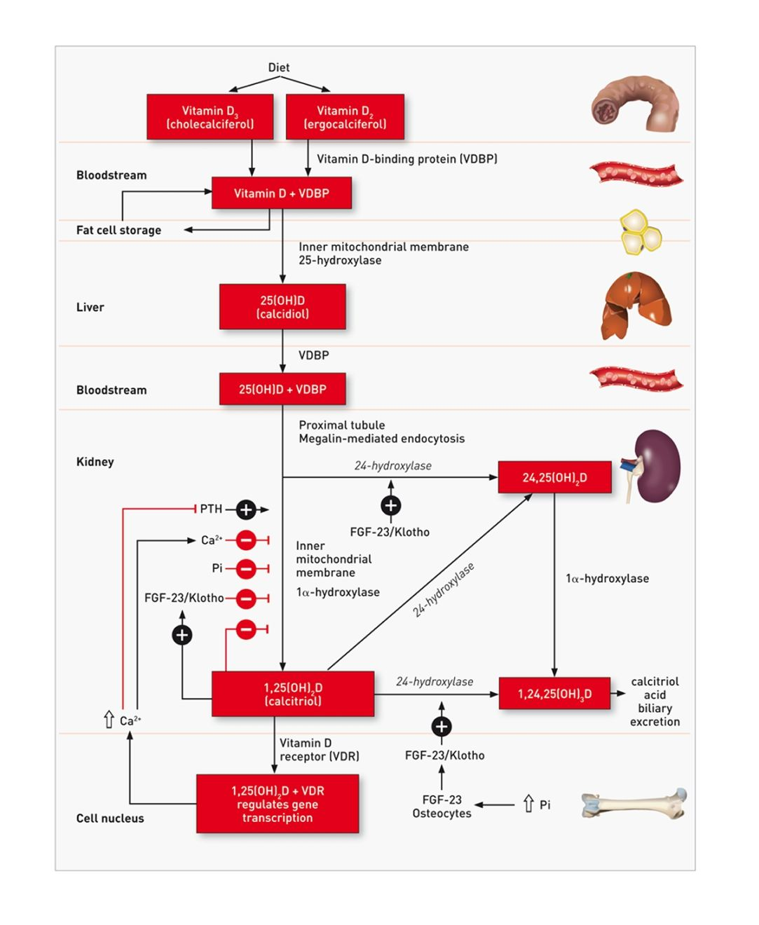
Kidney disease
Vitamin D metabolites have been measured in dogs with several forms of kidney disease, including acute renal failure, chronic kidney disease (CKD), and proteinuric kidney disease.Dogs with CKD have lower 25(OH)D and 1,25(OH)2 D concentrations compared with concentrations in control dogs [3] [4] [5]. Vitamin D metabolites are correlated with the stage of kidney disease (determined via International Renal Interest Society criteria), as indicated by the fact that concentrations of 25(OH)D, 1,25(OH)2 D and 24,25(OH)2 D are significantly decreased in dogs with stage 3 kidney disease, compared with control dogs [3] [4]. However, in other studies, many dogs had 25(OH)D and 1,25(OH)2 D concentrations within reference limits [6] [7]. One possible explanation for this lack of difference could be the inclusion of dogs with earlier stages of CKD. Alternatively, significant differences in concentrations of vitamin D metabolites may not have been detected because of relatively large reference ranges or the method used to calculate reference ranges.
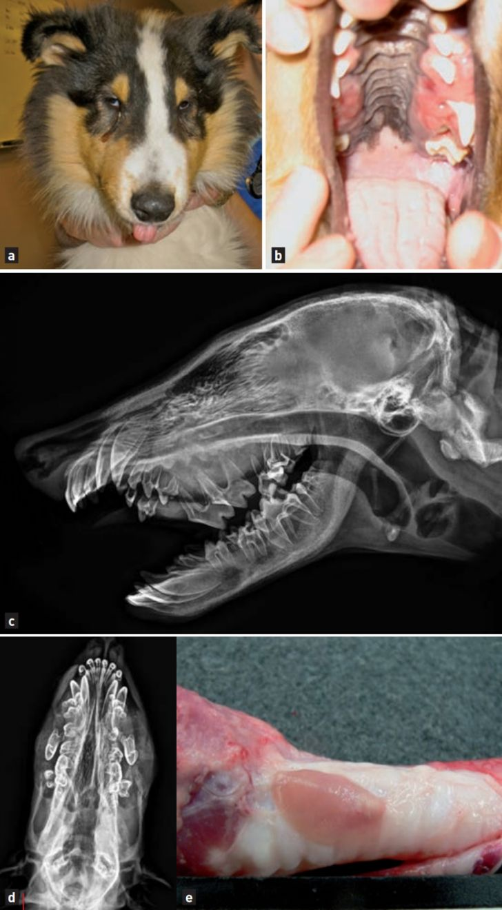
One of the consequences of CKD is the development of secondary hyperparathyroidism and CKD-induced mineral and bone disorders ( Figure 2 ). Plasma FGF23 concentrations are increased in dogs with CKD, and the concentration of FGF-23 has been found to be negatively correlated with 25(OH)D, 1,25(OH)2 D, and 24,25(OH)2 D concentrations and survival in dogs with CKD [4] [8].Calcitriol treatment has been recommended for several decades for dogs with CKD to reduce PTH concentrations and improve quality of life. However, prospective, controlled clinical studies are needed to determine the manner in which supplementation with various forms of vitamin D influences FGF-23 concentrations, Klotho expression, vitamin D repletion, quality of life, preservation of renal function, and survival.
Finally, dogs with acute renal failure have been reported to have significantly lower 25(OH)D and 1,25(OH)2 D concentrations, compared with control dogs, but most (7/10) of the dogs with acute renal failure had concentrations within reference limits [6]. These findings possibly could have been attributable to acute inflammation or critical illness, or could have been spurious results. Proteinuric dogs have significantly lower 25(OH)D, 1,25(OH)2 D, and 24,25(OH)2 D concentrations than control dogs. This relationship has been definitively established in people with proteinuria, and VDR activators are frequently prescribed to reduce proteinuria in such cases.
There are several mechanisms by which vitamin D metabolism can be disrupted with kidney disease, including decreased dietary intake of vitamin D, decreased enzymatic conversion from cholecalciferol to 25(OH)D in the liver, decreased activation via 1α-hydroxylase from 25(OH)D to 1,25(OH)2 D, and increased inactivation of 25(OH)D and 1,25(OH)2 D. With proteinuria, there are additional potential mechanisms to consider, including urinary loss of VDBP (with 25(OH)D and 1,25(OH)2 D bound to VDBP) and decreased endocytosis of 25(OH)D into renal cells because of decreased megalin expression in the proximal renal tubules. Furthermore, inflammation may act to reduce 25(OH)D concentrations.
Neoplasia
Decreased 25(OH)D concentrations have been linked to increased risk of numerous neoplasms in humans, and 1,25(OH)2 D has been found to have antineoplastic activity.Concentrations of circulating vitamin D metabolites have been measured in dogs with various tumors and serum 25(OH)D concentrations are significantly lower in many neoplastic conditions, including dogs with neoplasia and hemoabdomen, cutaneous mast cell tumor, and lymphoma. It is not clear whether dogs develop hypovitaminosis D secondary to neoplasia or whether hypovitaminosis D is actually a risk factor for development of cancer. Dogs with neoplasia are often ill; this puts them at risk of developing hypovitaminosis D from a reduced appetite, which leads to reduced cholecalciferol intake, and potentially from decreased intestinal absorption of cholecalciferol. It has recently been suggested that alteration of 25(OH)D concentrations in dogs with various neoplasms is mediated by ionized calcium concentrations [9].
Valerie J. Parker
Serum 1,25(OH)2 D concentrations have been measured in populations of dogs with lymphoma, both with and without hypercalcemia, with wide differences in findings. From an antineoplastic standpoint, calcitriol can have in vitro activity against osteosarcoma, squamous cell carcinoma, neoplastic prostatic epithelial cells, transitional cell carcinoma, mammary gland cancer, and mast cell tumor canine cell lines. One study revealed a synergistic effect of administering calcitriol with cisplatin against various tumors (e.g., osteosarcoma and chondrosarcoma) in dogs [10]. Investigators of another study found that calcitriol treatment could induce remission of mast cell tumors, but the trial was discontinued because of the high rate of toxicity (i.e., hypercalcemia and azotemia) observed [11].
Primary hyperparathyroidism
Although primary hyperparathyroidism is technically a neoplastic condition, it is separated here to avoid confusion with malignant conditions, because most dogs with primary hyperthyroidism have benign parathyroid gland adenomas. Compared with control dogs, five dogs with primary hyperparathyroidism had significantly lower serum 25(OH)D concentrations [7] although all values for the affected dogs were within reference limits. Serum 1,25(OH)2 D concentrations were significantly higher in dogs with primary hyperparathyroidism than in control dogs, and 1,25(OH)2 D concentrations in 4 of 5 dogs with primary hyperparathyroidism were above reference limits [7]. Both findings could possibly be attributed to an upregulating effect of PTH on renal 1α-hydroxylase activity, which would increase 1,25(OH)2 D synthesis.
In a study of 10 dogs with primary hyperparathyroidism treated by surgical excision of parathyroid gland adenomas, all had low 25(OH)D concentrations at the time of diagnosis, compared with control dogs, whereas 1,25(OH)2 D concentrations were within reference limits. At the time of the post-parathyroidectomy nadir in ionized calcium concentration, 25(OH)D concentrations did not differ from results at the time of initial diagnosis, but mean 1,25(OH)2 D concentrations were lower [12].
A diagnosis of primary hyperparathyroidism has traditionally been made on the basis of an increased ionized calcium concentration at the time of an inappropriately high concentration of PTH. The concentration of circulating 25(OH)D is an important regulatory factor for the suppression of PTH synthesis in humans (likely following its conversion to 1,25(OH)2 D within the parathyroid gland). Concentrations of PTH are higher in humans with concomitant lower circulating 25(OH) D concentrations. It is currently recommended that a diagnosis of primary hyperparathyroidism in humans is made only when 25(OH)D concentrations are sufficient or after 25(OH)D has been normalized following supplementation with vitamin D. The importance of concurrent evaluation of ionized calcium, PTH, and 25(OH)D concentrations to make an accurate diagnosis of primary hyperparathyroidism has not yet been investigated in veterinary medicine.
Gastrointestinal disease
Absorption of fat-soluble vitamins depends on adequate absorption of dietary fat; malabsorptive intestinal diseases can therefore adversely affect vitamin D absorption and ultimately contribute to hypovitaminosis D. Serum 25(OH)D and 1,25(OH)2 D concentrations have been evaluated in dogs with inflammatory bowel disease (IBD) and proteinlosing enteropathy (PLE), and both metabolites were significantly lower in the PLE group than in dogs with IBD or healthy dogs [13] [14]. Additionally, lower 25(OH)D concentrations were significantly correlated with duodenal inflammation and death [14] [15] [16].
It is possible that hypoalbuminemia contributes to hypovitaminosis D through loss of VDBP via diseased intestines. Alternatively, hypovitaminosis D may contribute to intestinal protein loss through the effect of vitamin D on the immune response. It is known that vitamin D receptor–knockout mice are more likely to develop induced IBD, and vitamin D-deficient diets predispose mice to colitis via dysregulated colonic antimicrobial activity and impaired homeostasis of enteric bacteria [17].
Orthopedic disease
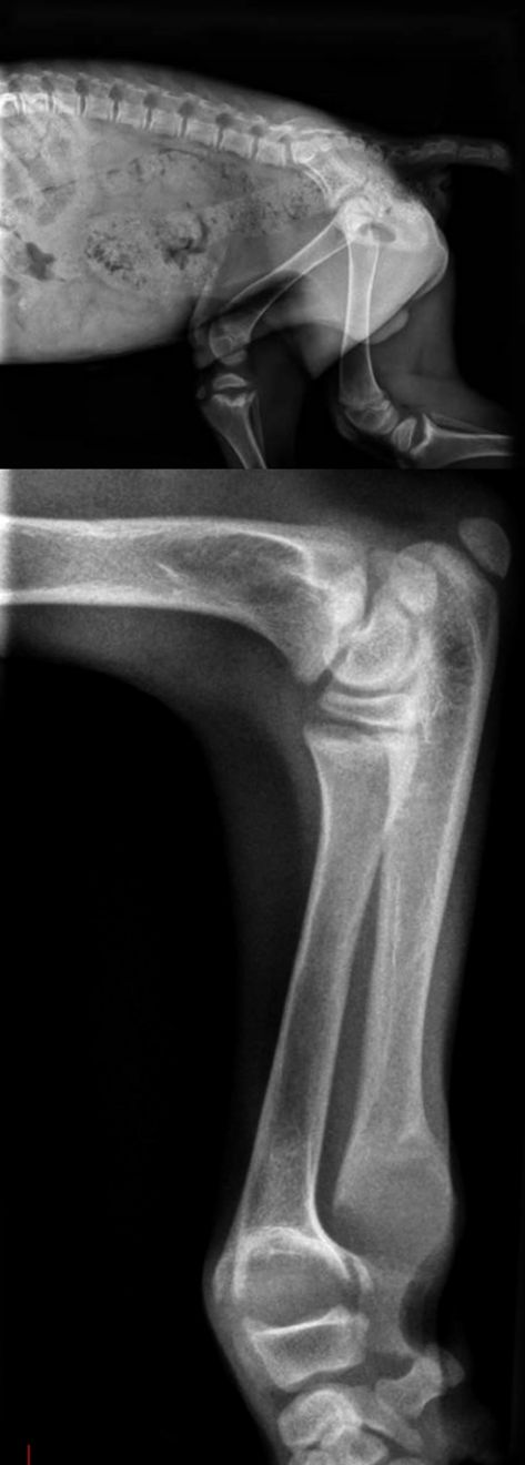
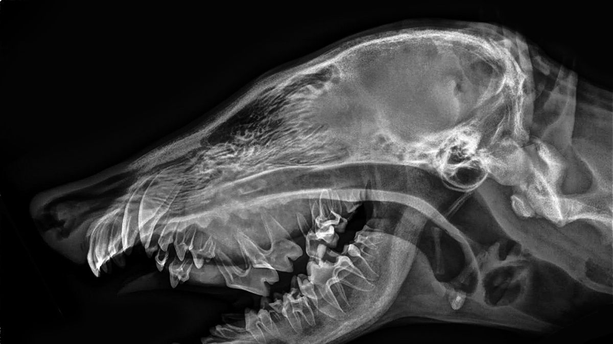
Osteoblasts and chondrocytes express 1α-hydroxylase and VDR but it is unknown whether vitamin D plays a direct or indirect role in bone growth and mineralization. Rickets is a metabolic bone disease typically caused by dietary deficiency of vitamin D, calcium or phosphorus, or by genetic defects affecting vitamin D or phosphorus metabolism ( Figure 3 ). The most common clinical abnormality is widening of the physeal growth plates of fast-growing bones such as the radius and ulna. Histologically, hypertrophic chondrocytes accumulate, which leads to thickened, irregular growth plates. Animals fed unbalanced meat-based diets without vitamin D supplementation are more likely to develop fibrous osteodystrophy, rather than rickets, because of the development of nutritional hyperparathyroidism. For an animal with dietaryinduced rickets, treatment entails transitioning the animal to a complete and balanced diet.
Two autosomal recessive disorders that cause vitamin D-dependent rickets (VDDR) in humans are recognized. Type I VDDR is caused by a defect in the gene encoding 1α-hydroxylase, which subsequently leads to inadequate activation of 25(OH)D to form 1,25(OH)2 D. This leads to 25(OH)D concentrations within the reference range but low 1,25(OH)2 D concentrations. Type II VDDR is caused by a defect in the VDR gene, which leads to hypocalcemia, secondary hyperparathyroidism, and high 1,25(OH)2 D concentrations. A few cases of both types of VDDR have been reported in dogs [18] [19]. Treatment of type I VDDR entails providing supplemental 1,25(OH)2 D and typically has a better prognosis than type II VDDR, which requires high doses of both 1,25(OH)2 D and calcium. Most mutations in people result in a defective VDR that can no longer respond to even high doses of 1,25(OH)2 D. Some children can be treated by high doses of 1,25(OH)2 D that overcome the defect in binding affinity for 1,25(OH)2 D.
Cardiovascular disease
Vitamin D plays a role in the pathophysiologic processes of cardiac disease. Cardiac myocytes express VDR and a calcitriol-dependent calciumbinding protein. In humans, hypovitaminosis D is associated with increased rates of myocardial infarction and cardiovascular events. An inverse relationship between vitamin D status and hypertension has been described in people, but a meta-analysis of 46 trials revealed that vitamin D supplementation had no effect on lowering blood pressure [20]. No study in dogs has documented a clear relationship between hypertension and vitamin D.The association between vitamin D and canine cardiac disease has been investigated. In one study that involved evaluation of 31 dogs with congestive heart failure, mean serum 25(OH)D concentrations were approximately 20% less than those of healthy control dogs [21]. Another study revealed that serum 25(OH)D concentrations were significantly lower in dogs with stage B2, C, or D chronic valvular disease (American College of Veterinary Internal Medicine criteria), compared with those in dogs with stage B1 chronic valvular disease (i.e., no evidence of cardiac remodeling). Serum 25(OH)D concentrations were significantly correlated with left ventricular and atrial sizes [22]. As with other diseases, decreased serum 25(OH)D concentrations may be linked to decreased dietary intake or increased inflammation. To the author’s knowledge, no veterinary studies have been conducted to evaluate FGF-23 or Klotho concentrations in relation to cardiovascular disease, although both FGF-23 and Klotho have been linked to cardiovascular disease (e.g., atherosclerosis, vascular stiffening, and left ventricular hypertrophy) in human CKD.
Inflammatory conditions
Vitamin D has been associated with inflammation and the immune system because most leukocytes express VDR. Serum 25(OH)D is a negative acutephase reactant and is typically inversely related to inflammatory markers (e.g., C-reactive protein, CRP) in humans. Furthermore, 25(OH)D and 1,25(OH)2 D modulate inflammation by inhibiting production of interleukin-6 and tumor necrosis factor-a. After strenuous racing, despite having higher CRP concentrations, sled dogs were found to have higher 25(OH)D concentrations [23]. No correlation between 25(OH)D and CRP concentrations in dogs with cancer has been noted [2]. With regard to leukocyte counts, serum 25(OH)D concentrations are significantly negatively correlated with neutrophil count, monocyte count, and interleukin-2 and -8 concentrations in dogs with chronic enteropathy [15].Other causes
Serum 25(OH)D concentrations have been investigated for some canine infectious diseases. Dogs with both neoplastic and non-neoplastic spirocercosis had significantly lower 25(OH)D concentrations than healthy dogs; dogs with neoplastic spirocercosis had significantly lower 25(OH)D concentrations than those with nonneoplastic spirocercosis [24].Granulomatous disease may induce hypercalcemia in dogs. This was originally thought to be mainly due to dysregulated production of calcitriol (i.e., increased production of 1,25(OH]2 D); however, there are granulomatous diseases in both humans and dogs in which hypercalcemia has been attributed to PTH-related peptide and not to calcitriol.Finally, dogs with acute polyradiculoneuritis have been shown to have lower 25(OH)D concentrations than dogs with idiopathic epilepsy [25]. The significance of this remains unknown.
Mortality rate and death
Low serum 25(OH)D concentrations have been linked to higher mortality rates in people, and serum 25(OH) D status has been shown to be predictive of 30-day mortality rate for hospitalized critically ill dogs [26]. Serum 25(OH)D concentration at time of diagnosis was a significant predictor of mortality rate for dogs with chronic enteropathy. It remains to be determined whether a low 25(OH)D concentration specifically influences the mortality rate, or if it is a consequence of increased inflammation and a greater severity of underlying disease.Vitamin D supplementation and toxicosis
Numerous studies have identified decreased concentrations of vitamin D metabolites in dogs with various diseases; however, it has not yet been determined whether such animals should receive supplemental vitamin D or vitamin D metabolites, and if so, the manner for providing them. Potential options include vitamin D2 (ergocalciferol), vitamin D3 (cholecalciferol), calcidiol, calcitriol, or other VDR activators (e.g., paricalcitol).
In a prospective study of canine atopic dermatitis, pruritus and lesion scores improved with cholecalciferol intake [1]. There was minimal toxicity observed but extremely high doses (up to 1400 IU/kg, higher than recommended by either AAFCO or NRC) were required to affect serum 25(OH)D concentrations and clinical signs. Recently, a modified-release formulation of 25(OH)D has been approved for the treatment of humans with advanced CKD4 . Providing supplemental 25(OH)D to dogs more rapidly and efficiently increases serum 25(OH)D concentrations than does cholecalciferol, but additional studies are necessary to elucidate appropriate dosing recommendations.
4 Rayaldee, OPKO Healthy Inc, Miami, Fla
The goal of supplementation with vitamin D or 25(OH)D should be to increase serum 25(OH)D concentrations and improve outcomes specific to the disease being managed (e.g., reducing pruritus or improving the survival rate or duration). The form of supplemental vitamin D administered, half-life of the product, and potential for toxic effects may differ, so caution must be exercised, and treated animals must be monitored closely.
Vitamin D toxicosis is most commonly diagnosed after the development of hypercalcemia and a subsequent risk for acute kidney injury and soft tissue mineralization. Development of hypercalcemia as a result of vitamin D toxicosis is a relatively late finding. Several factors influence the potential for vitamin D toxicosis, including lipophilicity, affinity of vitamin D metabolites for VDBP, and rates of metabolite synthesis and degradation. Vitamin D is fat soluble, a major reason why it has a long whole-body half-life of approximately 2 months. Half-lives for 25(OH)D and 1,25(OH)2 D are approximately 2-3 weeks and 4-6 hours, respectively.
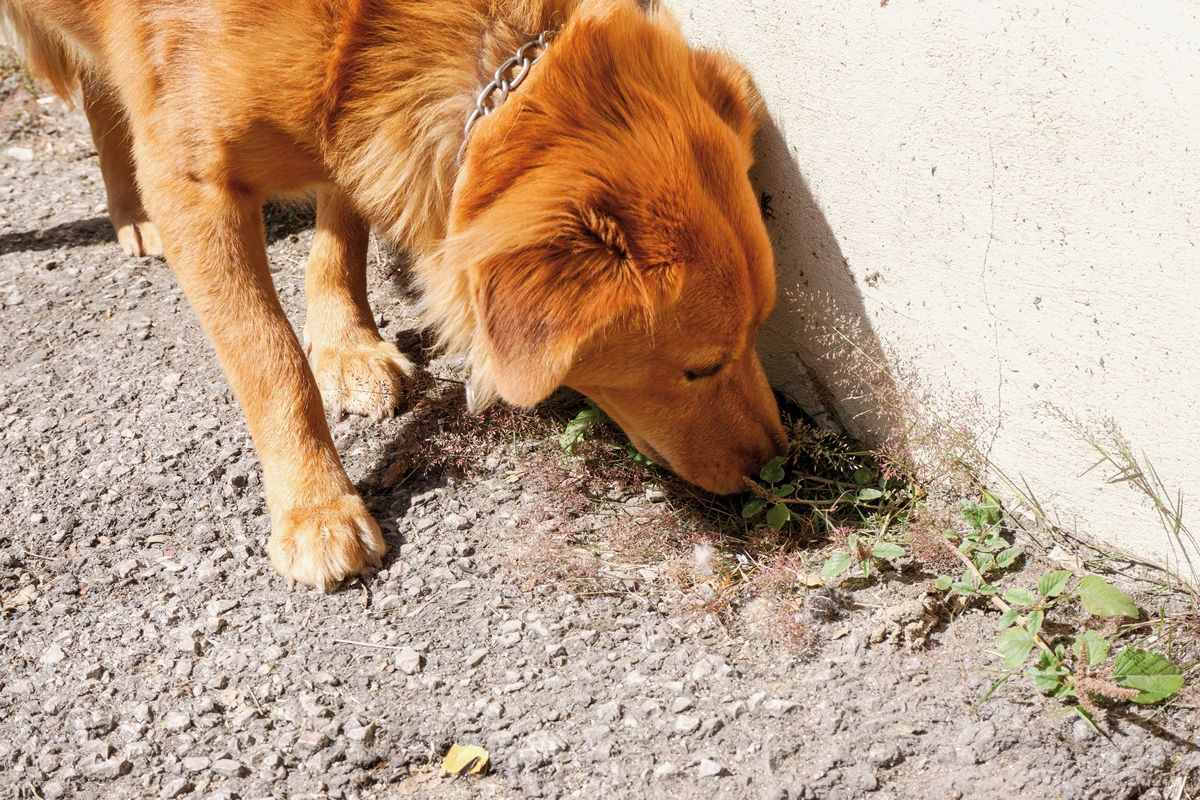
Vitamin D toxicosis in humans that results in hypercalcemia is thought to occur when serum 25(OH)D concentrations exceed 100-150 ng/mL. In studies of various animal species (rats, cows, pigs, rabbits, dogs, and horses), plasma 25(OH)D concentrations associated with hypercalcemia exceed 150 ng/mL. The most commonly encountered forms of vitamin D toxicosis in dogs include ingestion of cholecalciferol rodenticides ( Figure 4 ) and skin creams that contain calcitriol or an analogue (calcipotriol/calcipotriene). Occasionally, misformulation of commercial pet foods may contribute to vitamin D toxicosis. Iatrogenic toxicosis, typically determined by measurement of 1,25(OH)2 D concentrations, may occur secondary to provision of supplemental calcitriol for management of renal secondary hyperparathyroidism, primary hypoparathyroidism, PLE, or pre- or postsurgical treatment of primary hyperparathyroidism.
Note that hypercalciuria develops during the early phases of vitamin D toxicosis, before hypercalcemia develops, and can have a negative impact by increasing the risk of developing calciumcontaining uroliths and renal injury. The urinary calcium-to-creatinine ratio is used to detect hypercalciuria in humans, and this concept has received attention in the investigation of dogs that form calcium-containing uroliths.
Vitamin D homeostasis is characterized by complex interactions between vitamin D metabolites, ionized calcium, phosphorus, FGF-23, and Klotho, and regulatory pathways can be disrupted in a variety of ways. Although reference limits for serum vitamin D metabolites in healthy dogs remain to be determined, many diseases have been associated with lower concentrations of vitamin D metabolites, whereas some have been associated with increased concentrations. The chicken-and-egg conundrum often applies to these diseases, and it is not definitively clear whether vitamin D deficiency is the cause or the result of these diseases. Additional studies are needed to determine whether vitamin D supplementation for dogs with certain diseases would improve patient outcomes, and the form and dosing regimen that would best provide this supplemental vitamin D.

Valerie J. Parker
DVM, Dipl. ACVIM, Dipl. ACVNDr. Parker received her DVM from Tufts University and went on to complete a smallanimal internship at the Animal Medical Center in New York City. She then undertook asmall animal internal medicine residency at Iowa State University and a clinical nutritionresidency at Tufts University. She is currently associate professor at OSU, with researchinterests that include vitamin D status in chronic kidney disease and its relationship tonutrition management.
References
- Klinger CJ, Hobi S, Johansen C, et al. Vitamin D shows in vivo efficacy in a placebo-controlled, double-blinded, randomised clinical trial on canine atopic dermatitis. Vet Rec 2018;182:406.
- Selting KA, Sharp CR, Ringold R, et al. Serum 25-hydroxyvitamin D concentrations in dogs – correlation with health and cancer risk. Vet Comp Oncol 2016;14:295-305.
- Cortadellas O, Fernandez del Palacio MJ, Talavera J, et al. Calcium and phosphorus homeostasis in dogs with spontaneous chronic kidney disease at different stages of severity. J Vet Intern Med 2010;24:73-79.
- Parker VJ, Harjes LM, Dembek K, et al. Association of vitamin D metabolites with parathyroid hormone, fibroblast growth factor-23, calcium, and phosphorus in dogs with various stages of chronic kidney disease. J Vet Intern Med 2017;31:791-798.
- Galler A, Tran JL, Krammer-Lukas S, et al. Blood vitamin levels in dogs with chronic kidney disease. Vet J 2012;192:226-231.
- Gerber B, Hassig M, Reush CE. Serum concentrations of 1,25-dihydroxycholecalciferol and 25-hydroxycholecalciferol in clinically normal dogs and dogs with acute and chronic renal failure. Am J Vet Res 2003;64:1161-1166.
- Gerber B, Hauser B, Reusch CE. Serum levels of 25-hydroxycholecalciferol and 1,25-dihydroxycholecalciferol in dogs with hypercalcaemia. Vet Res Commun 2004;28:669-680.
- Rudinsky AJ, Harjes LM, Quimby J, et al. Relationship between fibroblast growth factor-23 and survival in dogs with chronic kidney disease. Submitted, J Vet Int Med 2018.
- Weidner N, Woods JP, Conlon P, et al. Influence of various factors on circulating 25(OH) vitamin D concentrations in dogs with cancer and healthy dogs. J Vet Intern Med 2017;31:1796-1803.
- Rassnick KM, Muindi JR, Johnson CS, et al. In vitro and in vivo evaluation of combined calcitriol and cisplatin in dogs with spontaneously occurring tumors. Cancer Chemother Pharmacol 2008;62:881-891.
- Malone EK, Rassnick KM, Wakshlag JJ, et al. Calcitriol (1,25-dihydroxycholecalciferol) enhances mast cell tumour chemotherapy and receptor tyrosine kinase inhibitor activity in vitro and has singleagent activity against spontaneously occurring canine mast cell tumours. Vet Comp Oncol 2010;8:209-220.
- Song J. Evaluation of parathyroid hormone and preoperative vitamin D as predictive factors for post-operative hypocalcemia in dogs with primary hyperparathyroidism. MS thesis, Dept. Vet Clinical Sciences, College of Veterinary Medicine, OSU 2016.
- Gow AG, Else R, Evans H, et al. Hypovitaminosis D in dogs with inflammatory bowel disease and hypoalbuminaemia. J Small Anim Pract 2011;52:411-418.
- Titmarsh H, Gow AG, Kilpatrick S, et al. Association of vitamin D status and clinical outcome in dogs with a chronic enteropathy. J Vet Intern Med 2015;29:1473-1478.
- Titmarsh HF, Gow AG, Kilpatrick S, et al. Low vitamin D status is associated with systemic and gastrointestinal inflammation in dogs with a chronic enteropathy. PLoS One 2015;10:e0137377.
- Allenspach K, Rizzo J, Jergens AE, et al. Hypovitaminosis D is associated with negative outcome in dogs with protein-losing enteropathy: a retrospective study of 43 cases. BMC Vet Res 2017;13:96.
- Lagishetty V, Misharin AV, Liu NQ, et al. Vitamin D deficiency in mice impairs colonic antibacterial activity and predisposes to colitis. Endocrinology 2010;151:2423-2432.
- Johnson KA, Church DB, Barton RJ, et al. Vitamin D-dependent rickets in a Saint Bernard dog. J Small Anim Pract 1988;29:657-666.
- LeVine DN, Zhou Y, Ghiloni RJ, et al. Hereditary 1,25-dihydroxyvitamin Dresistant rickets in a Pomeranian dog caused by a novel mutation in the vitamin D receptor gene. J Vet Intern Med 2009;23:1278-1283.
- Beveridge LA, Struthers AD, Khan F, et al. Effect of vitamin D supplementation on blood pressure: a systematic review and meta-analysis incorporating individual patient data. JAMA Intern Med 2015;175:745-754.
- Kraus MS, Rassnick KM, Wakshlag JJ, et al. Relation of vitamin D status to congestive heart failure and cardiovascular events in dogs. J Vet Intern Med 2014;28:109-115.
- Osuga T, Nakamura K, Morita T, et al. Vitamin D status in different stages of disease severity in dogs with chronic valvular heart disease. J Vet Intern Med 2015;29:1518-1523.
- Spoo JW, Downey RL, Griffitts C, et al. Plasma vitamin D metabolites and C-reactive protein in stage-stop racing endurance sled dogs. J Vet Intern Med 2015;29:519-525.
- Rosa CT, Schoeman JP, Berry JL, et al. Hypovitaminosis D in dogs with spirocercosis. J Vet Intern Med 2013;27:1159-1164.
- Laws EJ, Kathrani A, Harcourt-Brown TR, et al. 25-Hydroxyvitamin D3 serum concentration in dogs with acute polyradiculoneuritis compared to matched controls. J Small Anim Pract 2018;59:222-227.
- Jaffey JA, Backus RC, McDaniel KM, et al. Serum vitamin D concentrations in hospitalized critically ill dogs. PLoS One 2018;23:e0194062.