Introduction
Pododermatitis is the name given to the clinical presentation of an inflammation affecting the paws, although it can involve the skin of the paws, the interdigital spaces and/or the nail folds (paronychia). Pododermatitis can be the only abnormality detected, or it may be accompanied by other dermatologic or systemic clinical signs, so any cat with footpad lesions warrants a complete dermatological and physical examination. Remember that pododermatitis is a descriptive term, not a final diagnosis, and there is more than one differential diagnosis!
The overall prevalence of feline pododermatitis among dermatological diagnoses is low [1]. Diseases affecting the pads include, though are not limited to, eosinophilic granuloma, pemphigus foliaceous, mosquito bite hypersensitivity, metastatic adenocarcinoma (so called "lung-digit syndrome") and plasma cell pododermatitis.
Plasma cell pododermatitis
Etiology and clinical signs
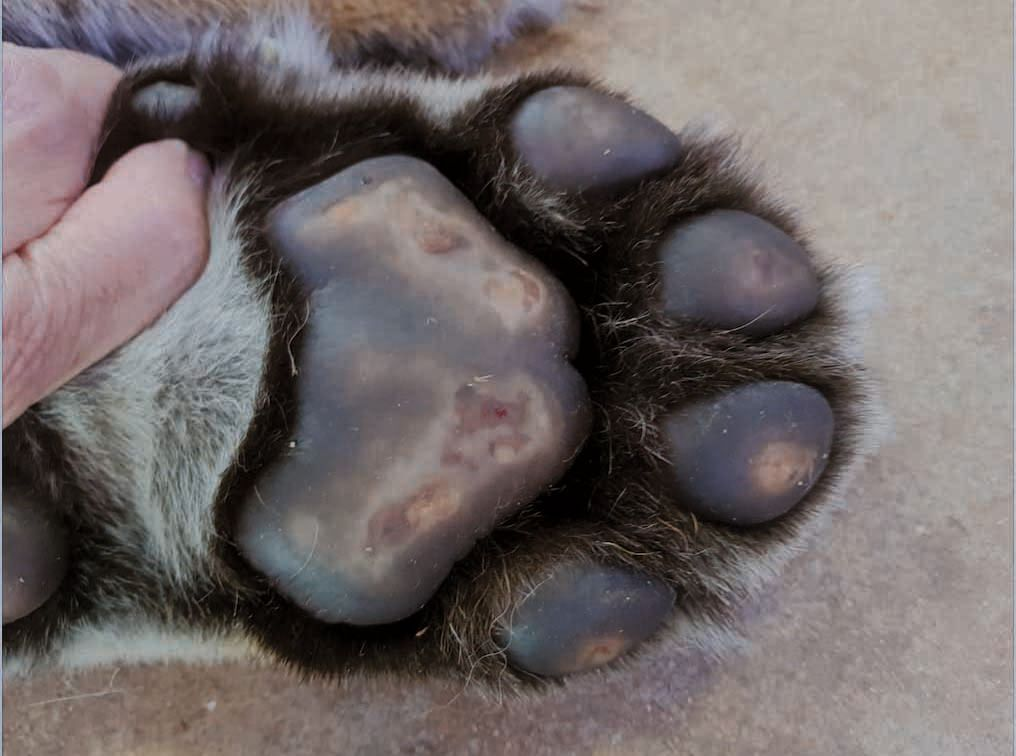
Plasma cell pododermatitis (PCP) is a rare dermatological condition of cats characterized by soft swelling of the paw-pads, which may eventually ulcerate to cause pain and lameness [2] [3] [4] [5] [6] [7]. The exact etiology and pathogenesis are not fully elucidated, although many studies have looked at possible triggers. A recent study [7] failed to detect several infectious agents (including Bartonella spp., Ehrlichia spp., Anaplasma phagocytophilum, Chlamydophila felis, Mycoplasma spp., Toxoplasma gondii, and feline herpesvirus) in tissue samples by immunohistochemistry and PCR. Previous publications reported concurrent FIV infection in 44-62% of cases, but the association is possibly incidental rather than causal [4] [5] [6] [7] [8]. An allergic etiology has also been suggested due to apparent seasonality [4] [8]. Regardless of cause, an immune-mediated pathogenesis purportedly underlies PCP, based both on lack of evidence of infectious agents and on the findings of tissue plasmacytosis, consistent hypergammaglobulinemia, and the favorable response to immunomodulating agents [2] [3].
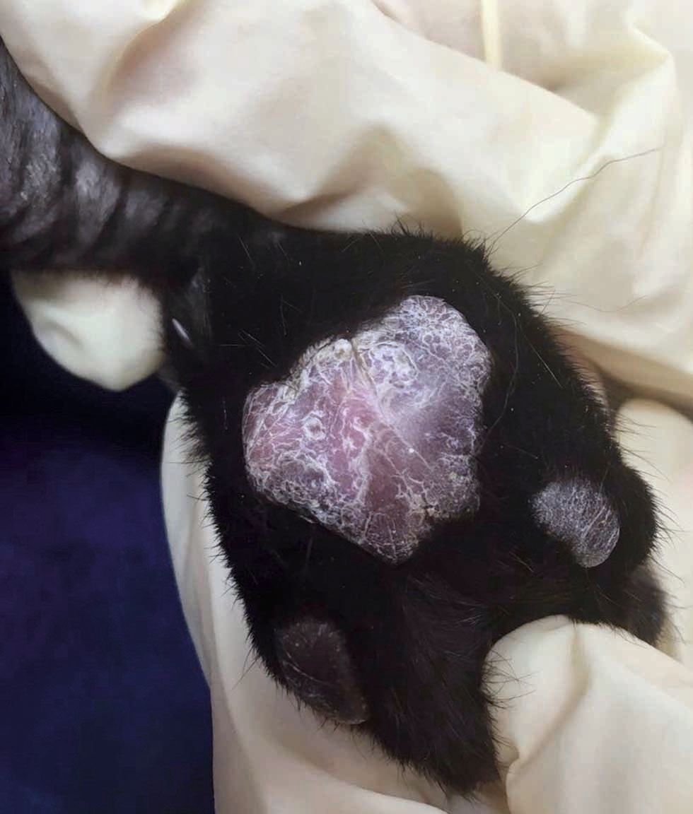
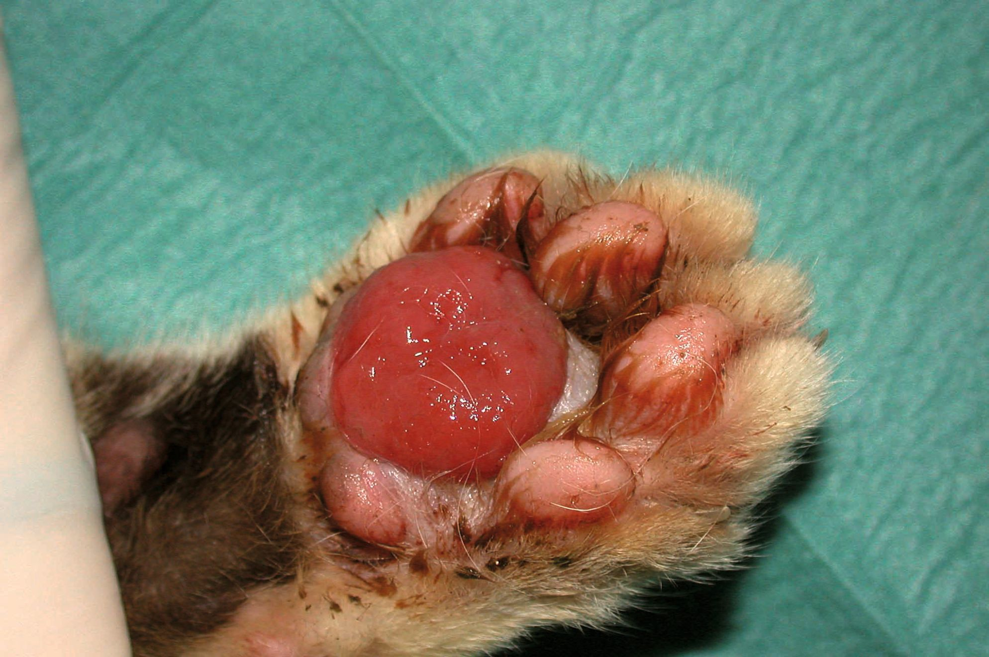
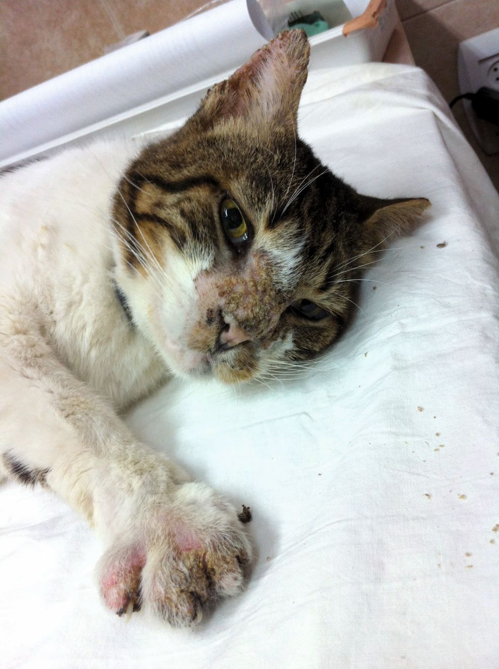
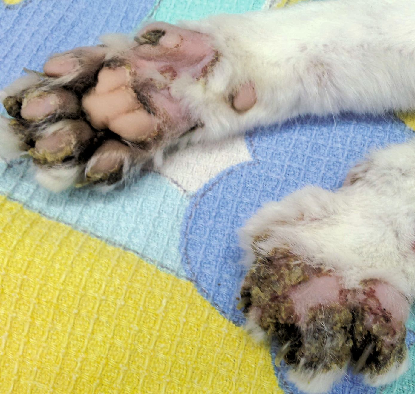
The disease can affect cats of almost any age, with no sex or breed predisposition. Clinical signs can range from painless soft, spongy swelling of the pads, with erythema, depigmentation, silvery striae and fine scaling, to ulceration and consequently bleeding, pain and lameness which can be severe (Figure 1) (Figure 2) (Figure 3). Typically, multiple paws are affected, with lesions predominantly on the central metacarpal and metatarsal pads. The digital pads may also be affected, but usually to a lesser degree. Rarely a single footpad will be affected. Secondary bacterial infection is not uncommon [2] [3] [4] [5] [6] [7] [8].
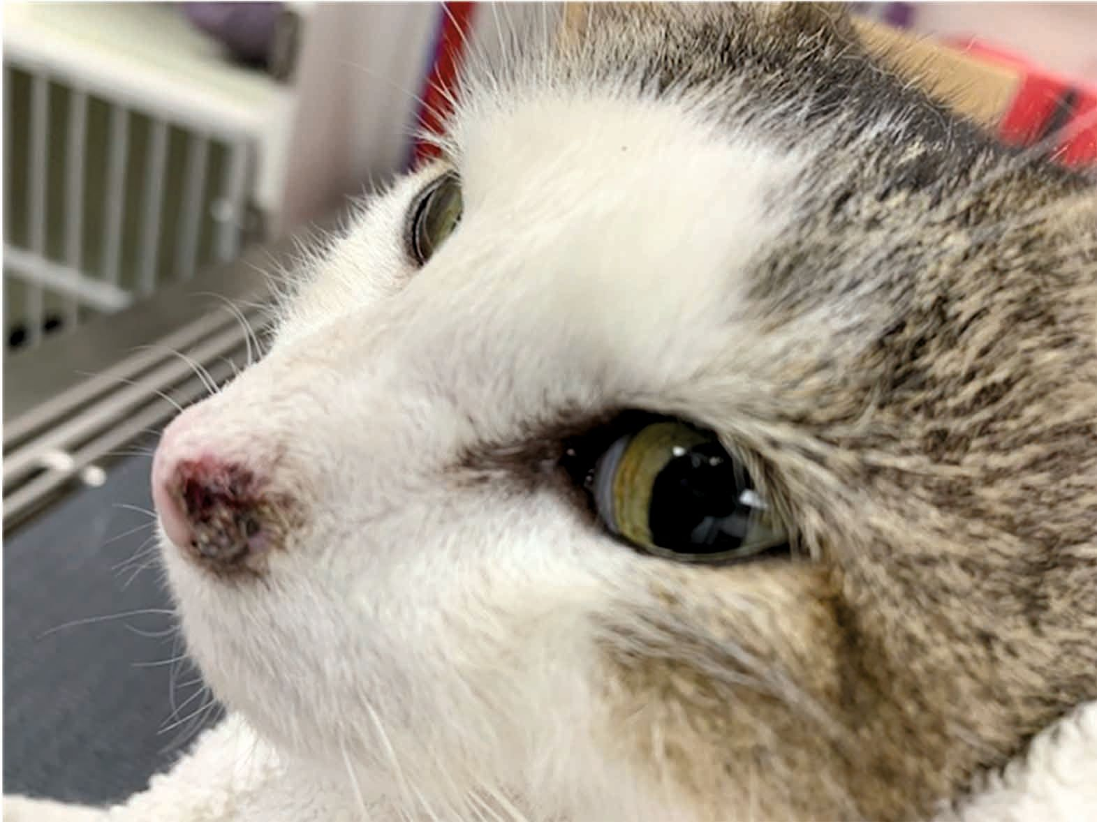
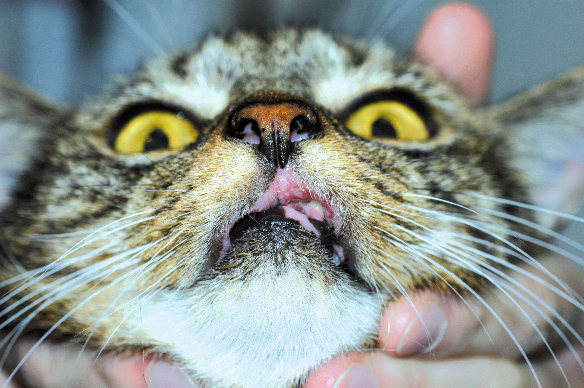
Some affected cats may show other clinical signs including poor body condition, hypersalivation [4], lymphadenopathy, pyrexia, anorexia, lethargy and (rarely) plasma cell dermatitis, with swelling of the nose or stomatitis [3] [4]. Immune-mediated glomerulonephritis or renal amyloidosis have also been reported [2] [3] [8]. Clinical pathologic findings may include anemia, leukocytosis and thrombocytopenia. A polyclonal gammopathy is present in all cases and can persist after treatment [2] [5].
Diagnosis
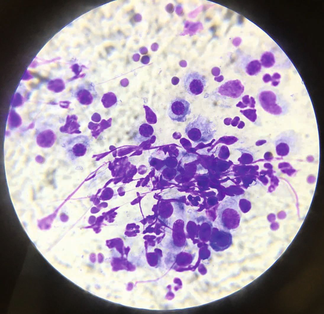
A tentative diagnosis can be made based on typical clinical signs, as the hallmark of PCP is soft swelling of the central pads, with or without ulceration, and usually in more than one foot. When this is present without other dermatological lesions the suspicion should be very high, and fine-needle aspiration (FNA) from a swollen pad showing plasma cells [2] [3] [6] (Figure 4) will support the notion. Definitive diagnosis is based on histopathology, but should avoid sampling ulcerated lesions. Histopathology features commonly include diffuse infiltration of plasma cells throughout the dermis and subcutis, and a variable number of lymphocytes, neutrophils and eosinophils in addition to Mott cells (plasma cells containing Russell bodies). Fibrosis and granulation tissue may be seen in chronic cases [2] [3] [4] [6] [7] [8]. The main differential diagnosis for PCP is eosinophilic granuloma, although the latter frequently has concurrent skin lesions, rarely affects multiple paws, and tends to affect the interdigital spaces rather than only the paw-pads [2] [3]. When a single pad is affected, neoplasia or a foreign body should be considered [3]. Multiple affected paws, especially when accompanied by paronychia, could suggest an infectious etiology.
Treatment
Plasma cell pododermatitis is characterized by a waxing and waning course, with some cases experiencing spontaneous remission, while others need life-long therapy [2] [3] [6] [8]. The current mainstay of treatment is immunomodulatory therapy and the initial therapy of choice is oral doxycycline [2] [3], an antibiotic with immunomodulatory properties. The recommended dose is 10 mg/kg q24H or 5 mg/kg q12H until clinical resolution, which might take up to 12 weeks [5] [6]. Since cats are prone to develop drug-induced esophagitis and subsequent esophageal stricture with this drug, it is imperative to administer the tablets or capsules with food and/or water [2] [3]. Cases with severe clinical signs and/or where doxycycline is ineffective will require systemic glucocorticoids: i.e., prednisolone at 2-4 mg/kg q24H, triamcinolone at 0.4-0.6 mg/kg q24H or dexamethasone 0.5 mg/kg, along with cyclosporine at 5-7.5 mg/kg q24H, to be tapered off slowly once in remission [2] [3]. Surgical excision has also been reported as curative, with no recurrence in the surgically treated pads after a follow-up period of 2 years [4] [8]. In cases requiring chronic treatment the immunomodulating drug should always be tapered down to the lowest frequency that allows the patient to remain comfortable.
Ronnie Kaufmann
Eosinophilic granuloma
Etiology and clinical signs
Feline eosinophilic granuloma (EG) affecting the paws is a manifestation of one of the three common presentations of feline eosinophilic granuloma complex (FEGC) [9] [10] [11]. Eosinophilic granuloma is a cutaneous reaction pattern and rarely a final diagnosis. Although the exact etiopathogenesis of FEGC is unclear, most evidence points to an underlying hypersensitivity to insects (mostly fleas), environmental allergens or foods. Some cases are idiopathic, although a genetic background may also be considered [9] [10] [11] [12].
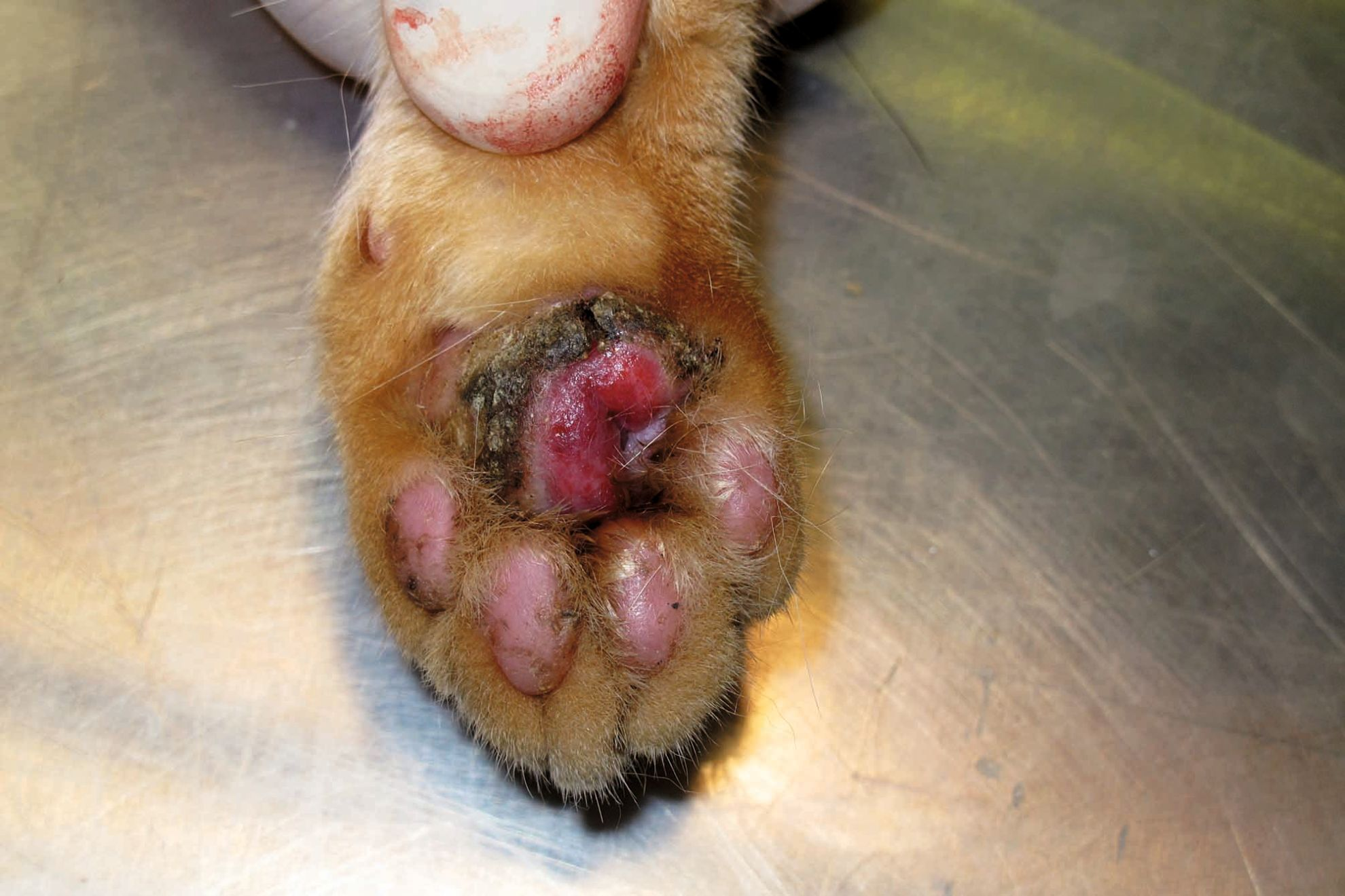
Excluding hereditary forms, which present at a younger age [9] [10] [12], no breed, sex or age predilection exists for EG [9] [10] [11]. The clinical presentation is very variable, as lesions can occur anywhere on the body, including the oral cavity. They are usually non-pruritic, well demarcated, raised to nodular, erythematous to orange-yellow in color, and occasionally ulcerated [9] [10] [11]. Lesions on the footpads are typically crusted and ulcerated (Figure 5). Eosinophilia may be present, although this is not a diagnostic feature [9] [10] [11]. A recent study has described atypical EG lesions affecting multiple paws in two littermates [12].
Diagnosis
When a cat is presented with crusty and/or ulcerated lesions of the paws a thorough dermatological examination should be performed, including the oral cavity, to exclude involvement of additional sites. Characteristic lesions in other locations should raise the suspicion of EG. Affected cats can also suffer simultaneously from other eosinophilic lesions and/or miliary dermatitis, further consolidating the suspicion of an eosinophilic disease [9] [10] [11]. Touch smears from ulcerated lesions or from under crusty lesions, or FNA from nodular lesions will aid diagnosis; samples usually show a mixed inflammatory reaction with a predominantly eosinophilic infiltrate [9] [10]. Tissue eosinophilia, albeit supportive, is not diagnostic in itself for EG, and histologic evaluation is required to establish a diagnosis [9] [10] [11] .
Differential diagnoses for EG footpad lesions include neoplasia (squamous cell carcinoma and mast cell tumor), infectious granuloma (bacterial folliculitis and furunculosis, mycobacteria, dermatophytes or deep fungal infection), abscessation, feline cowpox, foreign body reaction and sterile granulomatous disease [9] [10] [11]. Final diagnosis is achieved by histopathology, which is mandatory in solitary nodular lesions to rule out neoplasia or foreign bodies. Histopathology features consist of nodular to diffuse granulomatous dermatitis with a prominent eosinophilic infiltrate and multi-focal areas of collagen surrounded by degranulated eosinophils, known as “flame figures” [9] [10] [11]. Once the diagnosis of EG is confirmed, every attempt should be made to identify the underlying etiology.
Treatment
As with PCP, the prognosis for EG is variable, and spontaneous resolution is possible. In cases when an underlying cause is successfully identified and managed (i.e., flea or food hypersensitivity) the prognosis is very good as long as the offending triggers can be avoided. In idiopathic or atopy-related cases life-long treatment is necessary [9] [10] [11]. Therapy consists of immunomodulation, specifically oral prednisolone 1-2 mg/kg daily, then tapered to alternate-day dosing when possible. Sometimes higher doses (up to 4 mg/kg) may be required, and some cases will respond better to dexamethasone at 0.1-0.2 mg/kg (tapering to a maintenance dose of 0.05-0.1 mg/kg q72H), or triamcinolone 0.2-0.3 mg/kg daily. The goal is always to maintain the cat on the lowest possible dose and – more importantly – the lowest possible frequency that keeps it comfortable [9] [10] [11]. Every attempt should be made to avoid methylprednisolone acetate injections due to the higher risk of side effects, combined with the inability to withdraw treatment if adverse effects occur and the fact that increased doses may be required for cases with insufficient response [9]. Side effects of glucocorticoid therapy, though less common compared to dogs, include polydipsia, polyphagia, weight gain, diabetes mellitus, urinary tract infection, iatrogenic hyperadrenocorticism and feline cutaneous fragility syndrome, congestive heart failure, demodicosis and dermatophytosis [9] [10].
Cyclosporine at 7-7.5 mg/kg q24H has also proved efficacious for EG [9] [10] [11]. Due to a lag phase of 2-3 weeks, treatment should continue for at least 4 weeks and then taper to alternate days when possible, with some cases successfully managed on a q72H regimen. Side effects are not common, although 25% of cats may suffer transient gastrointestinal signs, including vomiting and diarrhea. In the author's experience co-administration of maropitant (2 mg/kg) during the first 2-3 weeks and/or gradually increasing the daily dose of cyclosporine can reduce the risk of vomiting. Other described adverse effects are weight loss, and (rarely) gingival hyperplasia, hypersalivation, anorexia and hepatic lipidosis [9] [10]. Cats prescribed cyclosporine should be FIV and FeLV negative and should not be allowed to hunt or eat raw meat, due to the risk of developing fatal toxoplasmosis.
Pemphigus foliaceus
Etiology and clinical signs
Pemphigus foliaceus (PF) is the most common autoimmune skin disease in cats, accounting for almost 1% of all cases seen by dermatologists [1]. It is a pustular, erosive and crusting dermatosis involving the face, ears and feet. Relapse is frequent and most cases will require long-term therapy, which should be personalized to each patient [13] [14] [15] [16] [17] [18].
Most cases appear to be idiopathic, with the remainder resulting from drug reactions and vaccines [13] [14] [15] [16], thymoma [14] [15] [16] and leishmaniasis [14]. The disease is characterized by autoantibodies produced against intercellular connections between epidermal cells in the superficial epidermis and follicular epithelium, known as desmosomes. These engender loss of cell-to-cell adhesion and the formation of acantholytic cells, which accumulate in sub-corneal and intraepidermal pustules which might eventually erupt into crusts, giving the disease its classy clinical crusty appearance [13] [14] [15] [16]. Though tissue-bound and circulating anti-keratinocyte IgG autoantibodies are detected in the majority of cats with PF, the exact pathomechanism is yet to be discovered, and the major target autoantigen in cats is still unknown [14] [15] [16] [17] [18].
The median age of onset is 6 years of age, with a range of 0.25-16 years [13] [14] [15] [16] [17] [18]. Domestic breed cats are over-represented, sex predilection has not been confirmed, but two recent reviews suggest that females may be marginally more prone to the condition [13] [14].
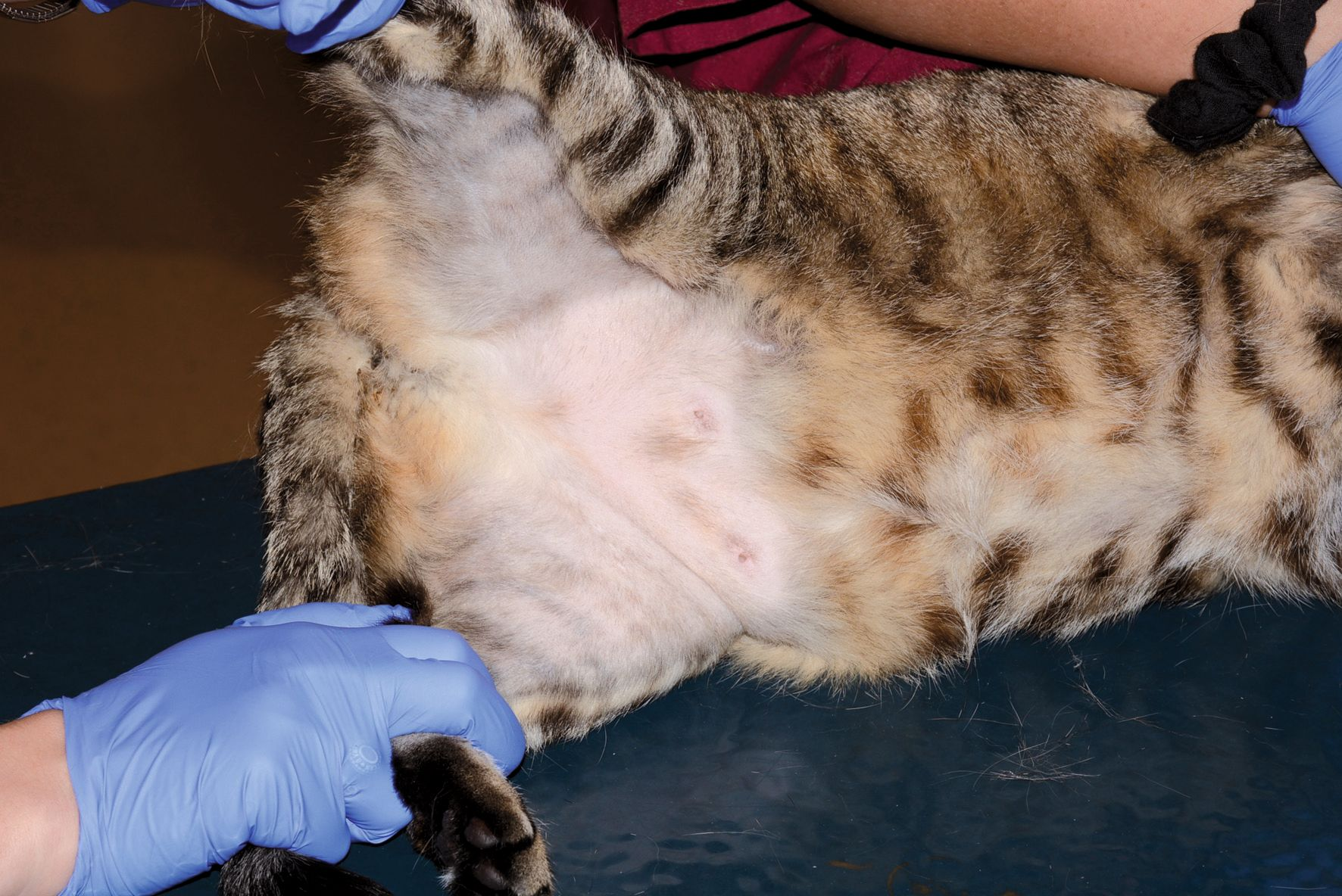
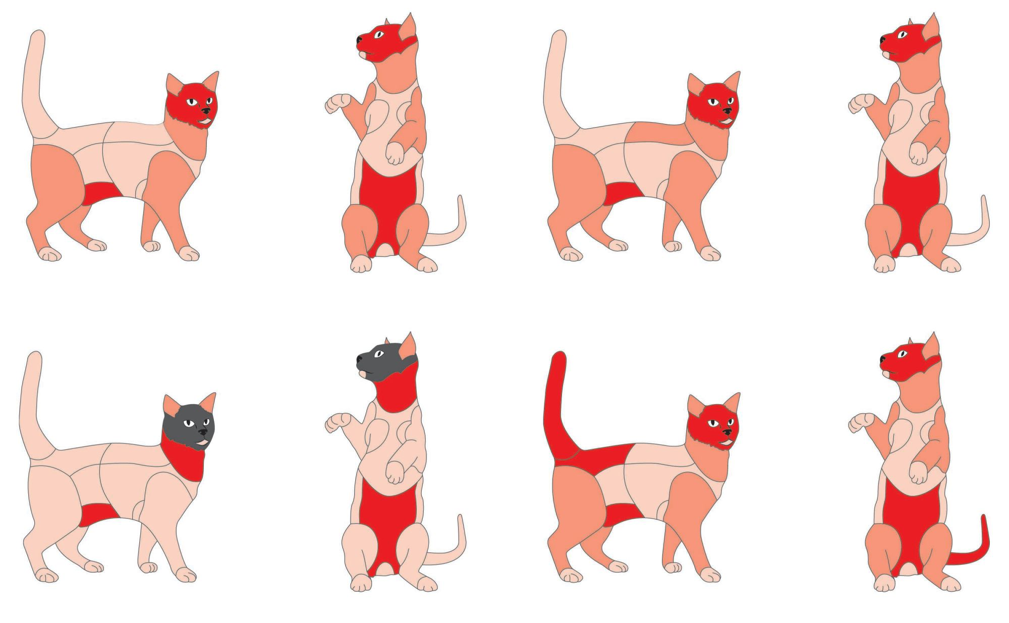
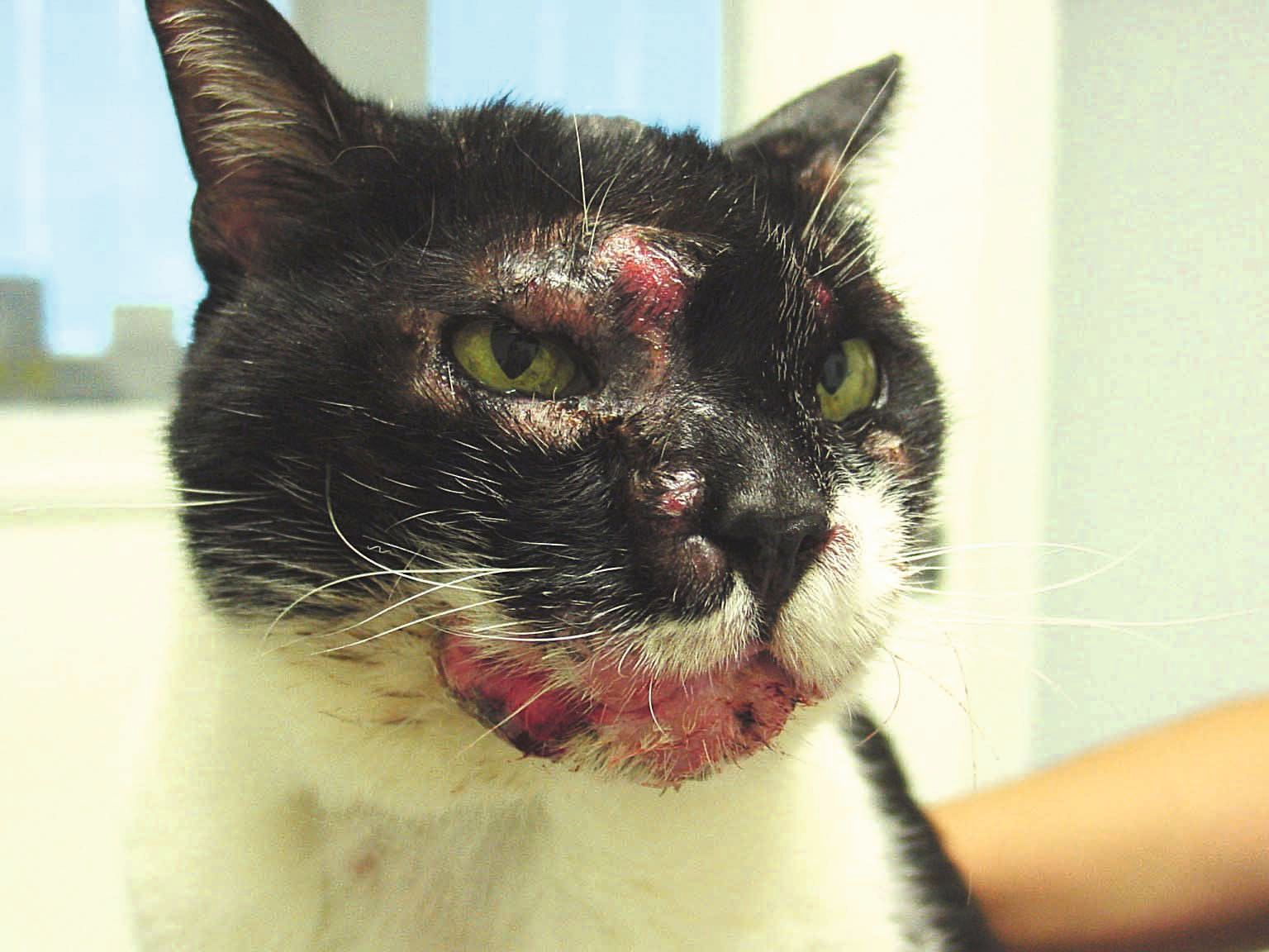
PF presents as a symmetric bilateral pustular disease, but since pustules rupture easily the typical clinical signs often include crusts, erosions, ulcerations, erythema and alopecia (Figure 6). In the majority of cases lesions involve more than one body region, most notably the head/face, claw folds and paws/pads [13] [14] [15] [16] [17] [18]. In some cases, claw folds can be the only affected area, which warrants consideration of PF in cats with erosive, exudative and/or crusting paronychia affecting the majority of the digits [14], which is sometimes so severe that the digits are forced to splay out (Figure 7). Pruritus is variable, and more than half of all cases will show systemic signs such as lethargy, pyrexia and anorexia [13] [14] [15] [16] [17] [18]. Lameness and pain are common if there are severe lesions affecting the paws. Clinicopathological findings of leukocytosis, neutrophilia, hyperglobulinemia and anemia are variably present [13] [15].
Diagnosis
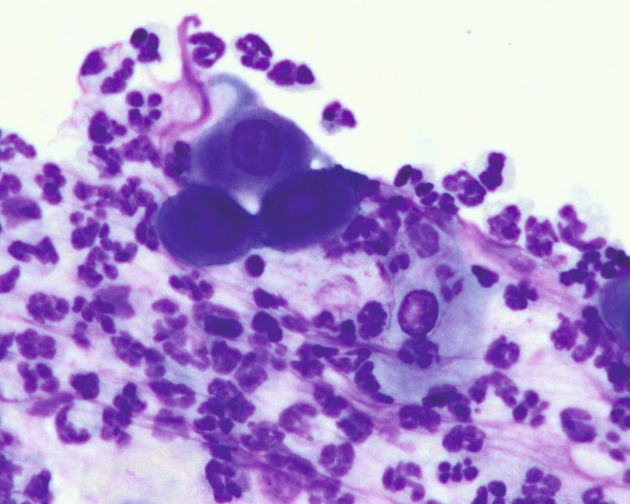
When typical clinical signs are present, most commonly pustules and crusts affecting multiple body sites with bilateral symmetry or multiple digits, cytology should be obtained directly from pustules or from underneath crusts. Characteristic findings include acantholytic cells (rounded keratinocytes with dark cytoplasm, resembling a “fried egg”) with intact neutrophils and occasionally eosinophils [15] [16] [17] [18] (Figure 8).
Definitive diagnosis is based on biopsies, either of whole pustules or (when a pustule cannot be sampled) crusts. Typical histopathological findings consist of crusting which frequently spans numerous hair follicles, sub-corneal or intragranular pustules, numerous acantholytic keratinocytes, and a predominantly neutrophilic dermal infiltrate which is often accompanied by eosinophils, mast cells and plasma cells [13] [14] [15] [16] [17] [18].
Treatment
Feline PF carries a favorable prognosis, with the majority of cats experiencing remission within a few weeks of treatment commencing [13] [14] [15] [16] [17] [18]. Importantly, disease control is defined as cessation of active lesions and healing of original lesions and not necessarily complete disappearance of all signs [14]. Most cats respond very well to glucocorticoid (GC) monotherapy, typically prednisolone at an initial dose of 2-4 mg/kg daily, with most documented cases responding to the lower dose. Other options include oral triamcinolone (0.2-0.6 mg/kg) or dexamethasone (0.1-0.2 mg/kg). Tapering the dose (20-25% reduction every 2-4 weeks) is recommended once the disease is inactive for at least 2 weeks and most original skin lesions have healed [14] [15] [17] [18].
Non-steroidal drugs reported to induce disease control in cats include cyclosporine (5-10 mg/kg daily) and chlorambucil (0.1-0.3 mg/kg daily) and are recommended in cases failing to respond to GC monotherapy, when severe side effects are seen with GC treatment, or when GC cannot be tapered off [14] [15] [16] [17] [18]. Most cats require long-term treatment due to the high frequency of relapse which can be due to drug tapering or discontinuation, but can also occur in well controlled cases [13] [14] [15] [16] [17] [18].
Owners should be aware of this latter possibility from the time of diagnosis, and discussion of different treatment protocols and side effects is warranted.
Ronnie Kaufmann
Mosquito bite hypersensitivity
Etiology and clinical signs
Feline mosquito bite hypersensitivity (MBH) is an uncommon, seasonal, pruritic dermatitis typically affecting the scarcely haired areas of the skin including the face, ears and paw-pads; the mosquito bite causes an IgE type I hypersensitivity reaction [10] [19].
There is no age, sex or breed predilection. Lesions are seen on poorly haired areas on the muzzle, pinnae, periaural and periorbital areas and the footpads, as cats will often lie with their legs outstretched. Erythematous papules or plaques progress to erosions and ulcers with crusts. Hyperkeratosis and variable pigmentation of the footpads is common, and pruritus can be intense. Occasionally regional lymphadenopathy, fever and mild eosinophilia are present [10] [19].
Diagnosis
The differential diagnosis will depend on the number of feet affected, and the aforementioned causes of pododermatitis should be considered. Cytology of lesions and lymph nodes can be supportive of MBH if dominated by eosinophils [19]. Typical histopathology findings consist of eosinophilic folliculitis and furunculosis, eosinophil exocytosis, a diffuse dermal eosinophilic inflammation and occasional flame figures [19]. Where the suspicion is high and there is a history of seasonality, mosquito avoidance will result in resolution within a few days.
Treatment
Usually a short course of systemic GC is needed to control acute clinical signs, but mosquito avoidance is the mainstay of therapy [10] [19]. Cats should be kept indoors during active mosquito hours and/or wear a protective anti-parasitic collar [19].
Metastatic adenocarcinoma
Etiology and clinical signs
Feline lung-digit syndrome (FLDS) describes an unusual pattern of metastasis from primary lung tumors, most commonly adenocarcinoma, to one or more digits [20] [21] [22]. Primary lung tumors are considered rare in the cat, with adenocarcinoma being the most common [21]. In one retrospective study, 88% of carcinomas in the digits represented metastases from primary lung carcinoma [23]. A more recent study looking at biopsies from 85 amputated feline digits identified a neoplastic disease in the majority (63) of cases, with 95.2% of them being malignant [24]. Metastatic adenocarcinomas of the lungs was the third most prevalent cause, which suggests that approximately 1 in 6 amputated digits submitted for histopathology in this study was a metastatic lesion [20]. The metastatic spread to the digits is thought to be due to the angioinvasive properties of these lesions and subsequent hematological spread. Cats have a high digital blood flow, offering a possible explanation for the significant rate of metastatic spread to this site [20].
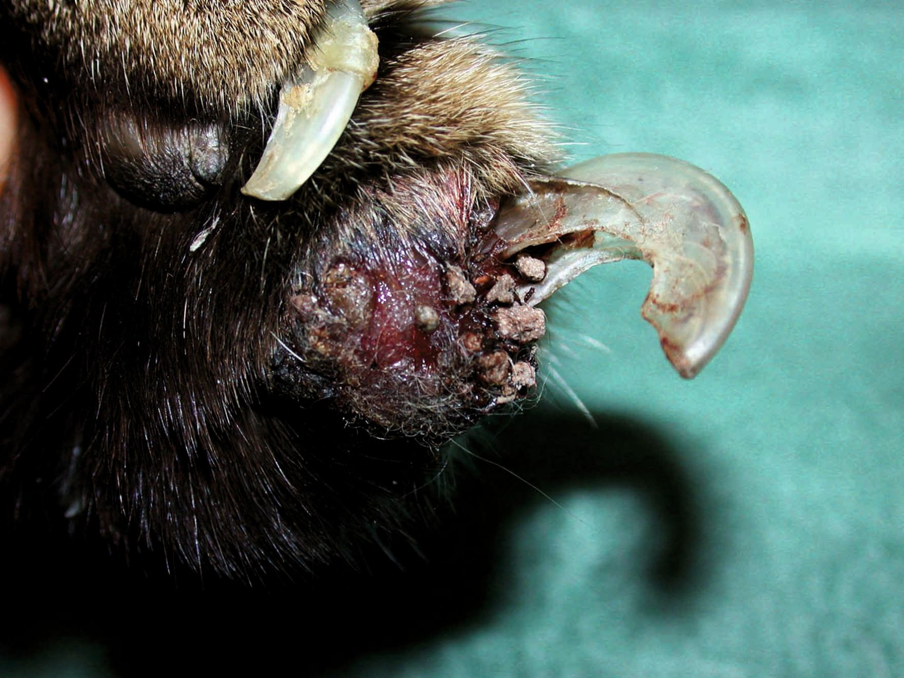
This is an uncommon disease affecting mostly older cats, with a mean age of 12 years (range 4-20 years) [20] [24]. Usually more than one digit is affected, with the exception of the dew claws [20] [22]. Typical signs include lameness, digital swelling, purulent nail bed discharge and fixed exsheathment of nails (Figure 9). Clinical signs associated with the primary lung tumor are often absent.
Diagnosis
Diagnosis can be easily confirmed by radiographs of the digits, which will classically show osteolysis of P3, potentially invading the P2-P3 intra-articular space, and possible osteolysis of P2; this is combined with thoracic radiography demonstrating a solitary circumscribed mass, usually in the caudal lung lobes [20] [21] [22]. Rarely, a thoracic radiograph will prove poorly sensitive, and a CT scan is recommended [22]. When in doubt, histopathology of an amputated digit will confirm the diagnosis [20] [22] [23] [24].
Treatment
The prognosis is grave, with reported survival time as low as 12 days and up to 125 days. Cats are usually euthanized due to pain, lethargy and anorexia [20] [21] and sudden death is possible [21]. Attempting surgical amputation should be carefully considered, given the poor prognosis [20] [21] [22].
Conclusion
Any cat presenting with paw-pad lesions should undergo a full dermatological and physical examination. When signs are confined to the feet, PCP and FLDS are the main differentials, and to a lesser degree EG, which occasionally only affects the paws; these can be tentatively differentiated by their gross appearance, supported by FNA findings. PF can also infrequently be confined to the paws, usually involving all feet and accompanied by severe paronychia. EG, MBH and PF more typically affect other body sites, but PF will usually have a bilateral symmetry with pustules and crusts, as opposed to the sporadic distribution of EG or localization of MBH to sparsely haired areas of the face. Lastly, systemic signs are more common in PF. A definitive diagnosis relies on histopathology, and although therapy commonly involves immunomodulation, it is different for every disease, as is the prognosis.

Ronnie Kaufmann
BSc., DVM, Dip. ECVD
Dr. Kaufmann received her veterinary degree from the Hebrew University of Jerusalem in 2006 and worked in general practice for three years before returning to the Koret School of Veterinary Medicine as a dermatology resident. She then remained as a clinician and lecturer at the school before being promoted to her current position as head of dermatology in 2017. Her main areas of interest include straelensiosis in companion animals and genetic dermatological conditions.
References
- Scott DW, Miller WH, Erb HN. Feline dermatology at Cornell University: 1407 cases (1988-2003). J Feline Med Surg 2012;15(4):307-316.
- Cain CL, Mauldin EA. Diagnostically Challenging Dermatoses of Cats. In: Little SE (ed.) August's Consultations in Feline Internal Medicine Vol. 7. 1st ed. St Louis, MO; WB Saunders 2016;295-306.
- Banovic F. Immune-Mediated Diseases. In: Noli C, Colombo S (eds.) Feline Dermatology. 1st ed. Switzerland AG, Springer Nature 2020;511-530.
- Guaguere E, Prelaud P, Degorce‐Rubiales F, et al. Feline plasma cell pododermatitis: a retrospective study of 26 cases. Vet Dermatol 2004;15:27.
- Scarampella F, Ordeix L. Doxycycline therapy in 10 cases of feline plasma cell pododermatitis: clinical, haematological and serological evaluations. Vet Dermatol 2004;15:27.
- Bettenay SV, Mueller RS, Dow K, et al. Prospective study of the treatment of feline plasmacytic pododermatitis with doxycycline. Vet Rec 2003;152:564-566.
- Bettenay SV, Lappin MR, Mueller RS. An immunohistochemical and polymerase chain reaction evaluation of feline plasmacytic pododermatitis. Vet Pathol 2007;44:80-83.
- Pereira PD, Faustino AMR. Feline plasma cell pododermatitis: a study of 8 cases. Vet Dermatol 2003;14:333-337.
- Buckley L, Nuttall T. Feline eosinophilic granuloma complexities: some clinical clarification. J Feline Med Surg 2012;14:471-481.
- Bloom PB. Canine and feline eosinophilic skin diseases. Vet Clin North Am Small Anim Pract 2006;36:141-160.
- Miller WH, Griffin CE, Campbell KL. Miscellaneous skin diseases. In: Muller and Kirk's Small Animal Dermatology. 7th ed. Philadelphia, WB Saunders, 2013;695-723.
- Pressanti C, Cadiergues MC. Feline familial pedal eosinophilic dermatosis in two littermates. J Feline Med Surg 2015;1(1):2055116915579683.
- Jordan TJM, Affolter VK, Outerbridge CA, et al. Clinicopathological findings and clinical outcomes in 49 cases of feline pemphigus foliaceus examined in Northern California, USA (1987-2017). Vet Dermatol 2019;30:209-219.
- Bizikova P, Burrows A. Feline pemphigus foliaceus: original case series and a comprehensive literature review. BMC Vet Res 2019;22:1-15.
- Preziosi DE. Feline pemphigus folliaceus. Vet Clin Small Anim 2019;49:95-104.
- Olivry T. A review of autoimmune skin diseases in domestic animals: 1 – superficial pemphigus. Vet Dermatol 2006;17:291-305.
- Bizikova P. Autoimmune Diseases. In: Noli C, Colombo S (eds.) Feline Dermatology. 1st ed. Switzerland AG, Springer Nature 2020;495-509.
- Klinger CJ, Mueller RS. Pemphigus foliaceus in feline patients. Vet Focus 2018;28.1:15-19.
- Mason K. Mosquito-byte Hypersensitivity. In: Noli C, Colombo S (eds.) Feline Dermatology. 1st ed. Switzerland AG Springer Nature, 2020;489-494.
- Goldfinch N, Argyle D. Feline lung-digit syndrome: unusual metastatic patterns of primary lung tumors in cats. J Feline Med Surg 2012;14:202-208.
- Sugiyama H, Maruo T, Shida T, et al. Clinical findings in lung-digit syndrome in five cats. J Jpn Vet Cancer Soc 2010;1(1):8-13.
- Thrift E, Greenwell C, Turner AL, et al. Metastatic pulmonary carcinomas in cats (“feline lung-digit syndrome”): further variations on a theme. J Feline Med Surg open rep 2017;1-8.
- van der Limde-Sipman JS, van den Ingh TS. Primary and metastatic carcinomas in the digits of cats. Vet Q 2010;3:141-145.
- Wobeser BK, Kidney BA, Powers BE, et al. Diagnoses and clinical outcomes associated with surgically amputated feline digits submitted to multiple veterinary diagnostic laboratories. Vet Pathol 2007;44:362-365.