Introduction
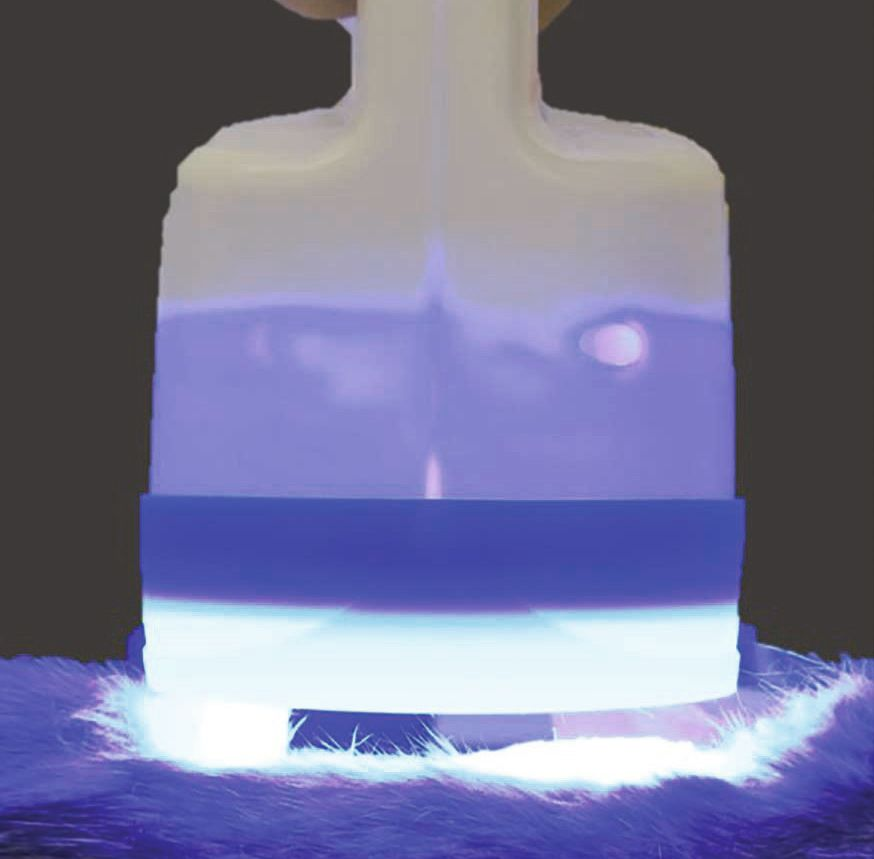
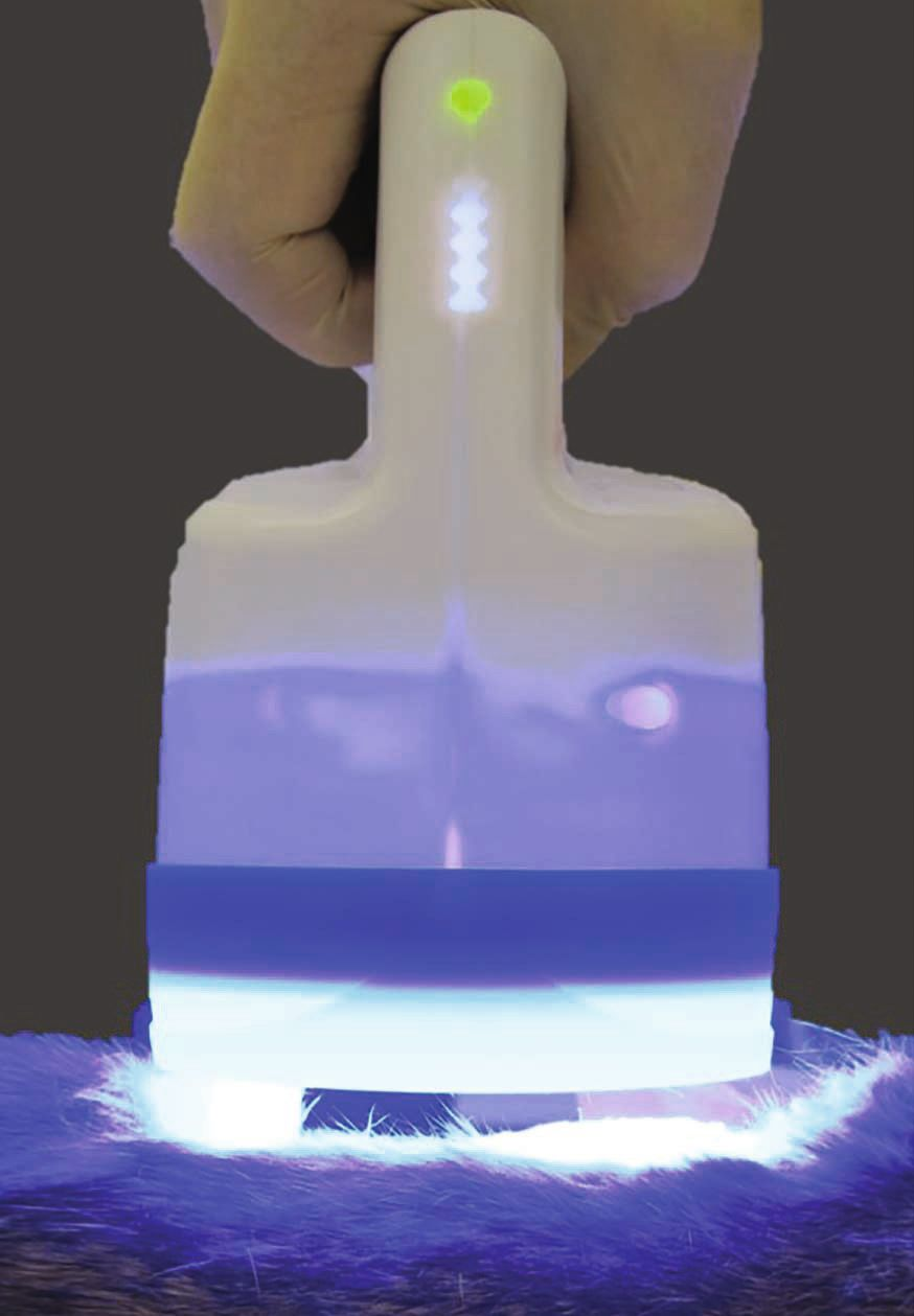
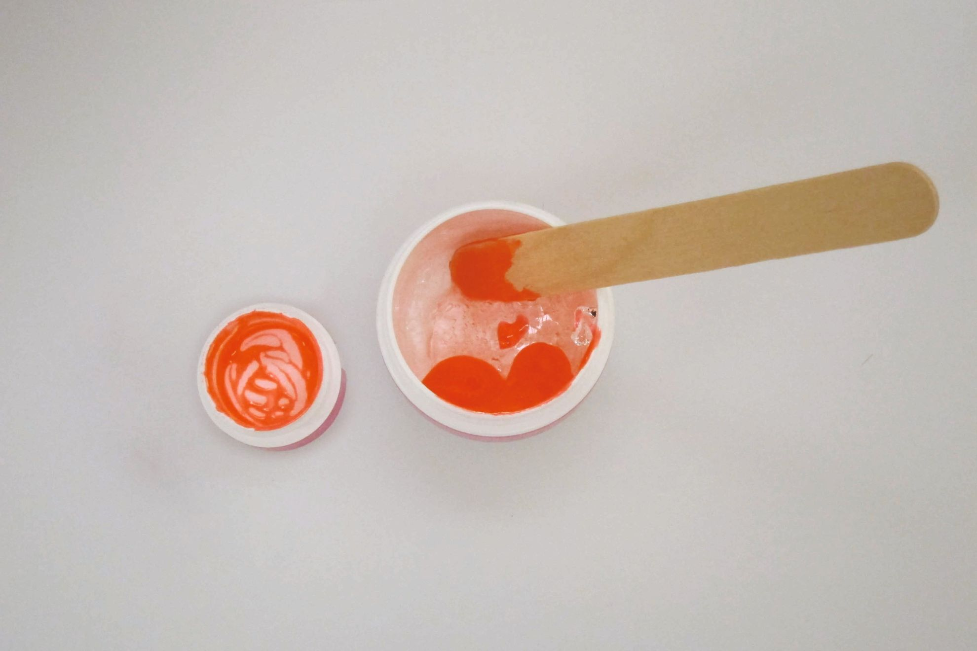
Fluorescence biomodulation (FBM) is a form of low-level laser therapy (LLLT) that is now gaining in popularity as a viable option for treating certain conditions in veterinary medicine. In general, LLLT methods typically employ photons of various wavelengths and operate at non-thermal irradiance levels to influence biological processes [1]. To do this, light must be able to penetrate tissues, with the depth of penetration being dictated by the light’s wavelength. The skin contains a variety of endogenous chromophores (molecules that absorb photons at certain wavelengths), the most common being hemoglobin and melanin; each has distinctive scattering and absorption coefficients that are significantly dependent on the wavelength of light [2]. Therefore, the choice of wavelength is considered one of the most important factors in LLLT. With a FBM system, a blue LED light (Figure 1) is used to activate a substrate, a photoconverting gel containing chromophores (Figure 2). The activated chromophores release energy in the form of fluorescent light which penetrates the patient’s skin; the depth of penetration varies depending on the emitted spectral profile [2]. Light in the blue-to-green spectrum has a penetration depth of approximately 1-2.5 mm into the skin, primarily affecting the epidermis and the upper layer of the dermis. Red light can penetrate nearly 5 mm into the skin, reaching deep into the dermis, and possibly even the panniculus [2]. This short article provides an insight into the use of FBM therapy for some skin conditions in clinical practice.
FBM in veterinary medicine
FBM was originally utilized in human medicine and is being now employed in veterinary medicine to treat various dermatological disorders, which are of course one of the most common reasons for pet owners to seek veterinary advice [3]. At least one commercially manufactured system is now available for use in veterinary practice in many countries. The treatment duration is short, non-painful and can usually be performed without sedation. The process is simple; the area requiring treatment is first clipped (if necessary) and then cleaned with sterile saline before the chromophore gel is applied to a thickness of approximately 2 mm with a spatula. This is then illuminated with the LED lamp for two minutes. Studies evaluating FBM have demonstrated the downregulation of pro-inflammatory cytokines and upregulation of anti-inflammatory cytokines. Growth factors that are important for proliferation of new granulation tissue, angiogenesis, and collagen remodeling – which together promote full wound healing – have been shown in studies to be elevated following treatment [1],[2],[4].
Pyoderma
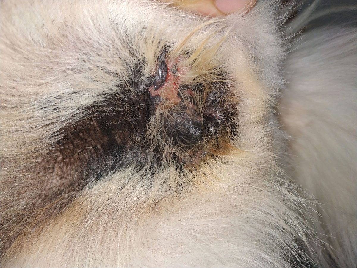
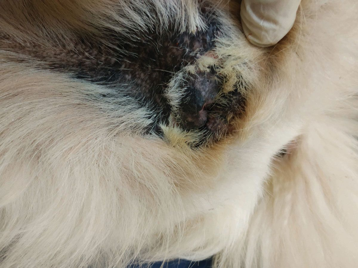
FBM has been applied with promising results to treat both superficial bacterial folliculitis [5] and deep otitis externa in dogs [6]. These studies show the technique is safe and can be used in conjunction with systemic antibiotics to accelerate the treatment of canine pyoderma. For superficial canine pyoderma, FBM could be considered for use as a sole treatment [1],[2],[5],[7], although further studies are needed to prove this suggestion. Figure 3 shows a dog with pyoderma of the vulva fold before treatment commenced. The pyoderma was treated with FBM at weekly intervals (with two consecutive applications per session) for two months, and the beneficial effects can be seen in Figure 4.
A prospective, randomized, and blinded clinical study assessed the efficacy of a FBM system to treat dogs with interdigital pyoderma. 36 dogs were randomly allocated to treatment groups of either antibiotic alone or antibiotic plus FBM application, with dogs in the latter group receiving the light treatment for two minutes twice a week until clinical resolution. Dogs were scored over a 12-week period, and the mean time-to-resolution of lesions was 4.3 weeks for those receiving both forms of therapy, as compared to 10.4 weeks for dogs that received only antibiotics. The conclusion was that FBM significantly reduced the time needed for clinical resolution [8]. FBM twice weekly can, therefore, be recommended as a therapeutic approach for managing interdigital pyoderma in dogs [9]; treating affected dogs once weekly (with two consecutive applications in the same session) could also be considered as an option [10], but larger randomized studies are needed to validate the existing suggestive data [9].
While these results show the clinical antibacterial efficacy of FBM, the mechanism remains unknown; two preliminary in-vitro studies were unable to demonstrate bactericidal activity of FBM [11] or blue LED light [12].
Otitis externa
A randomized non-blinded clinical trial suggested that FBM could also be beneficial in the treatment of otitis externa in dogs [13], a common condition accounting for up to 20% of consultations in small animal practice [14]. Application of FBM in the external ear canal may modulate inflammation, pain and bacterial growth [13] – although as mentioned above, in-vitro studies have not been able to determine why FBM or blue LED light should have bactericidal activity. However, in contrast, a preliminary in-vitro study has shown that FBM can inhibit the growth of Malassezia pachydermatis after at least four minutes of exposure [15].
Miscellaneous skin conditions
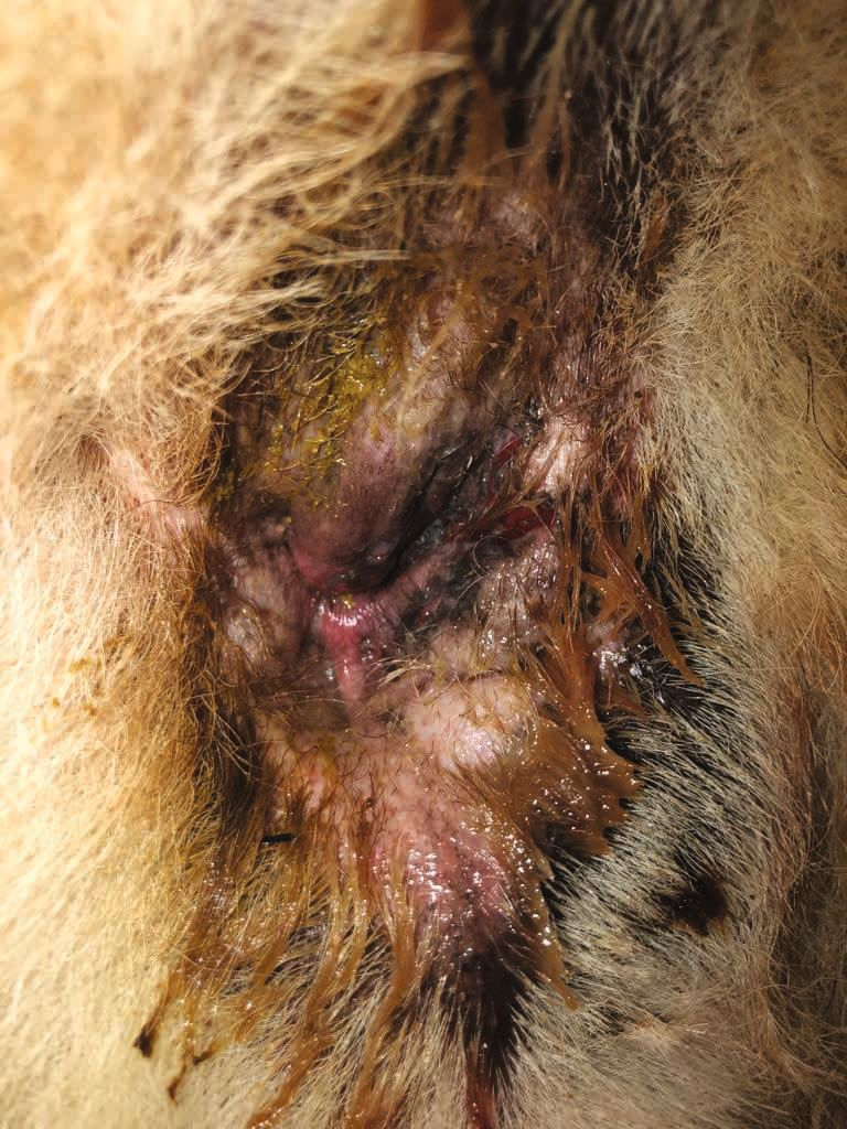
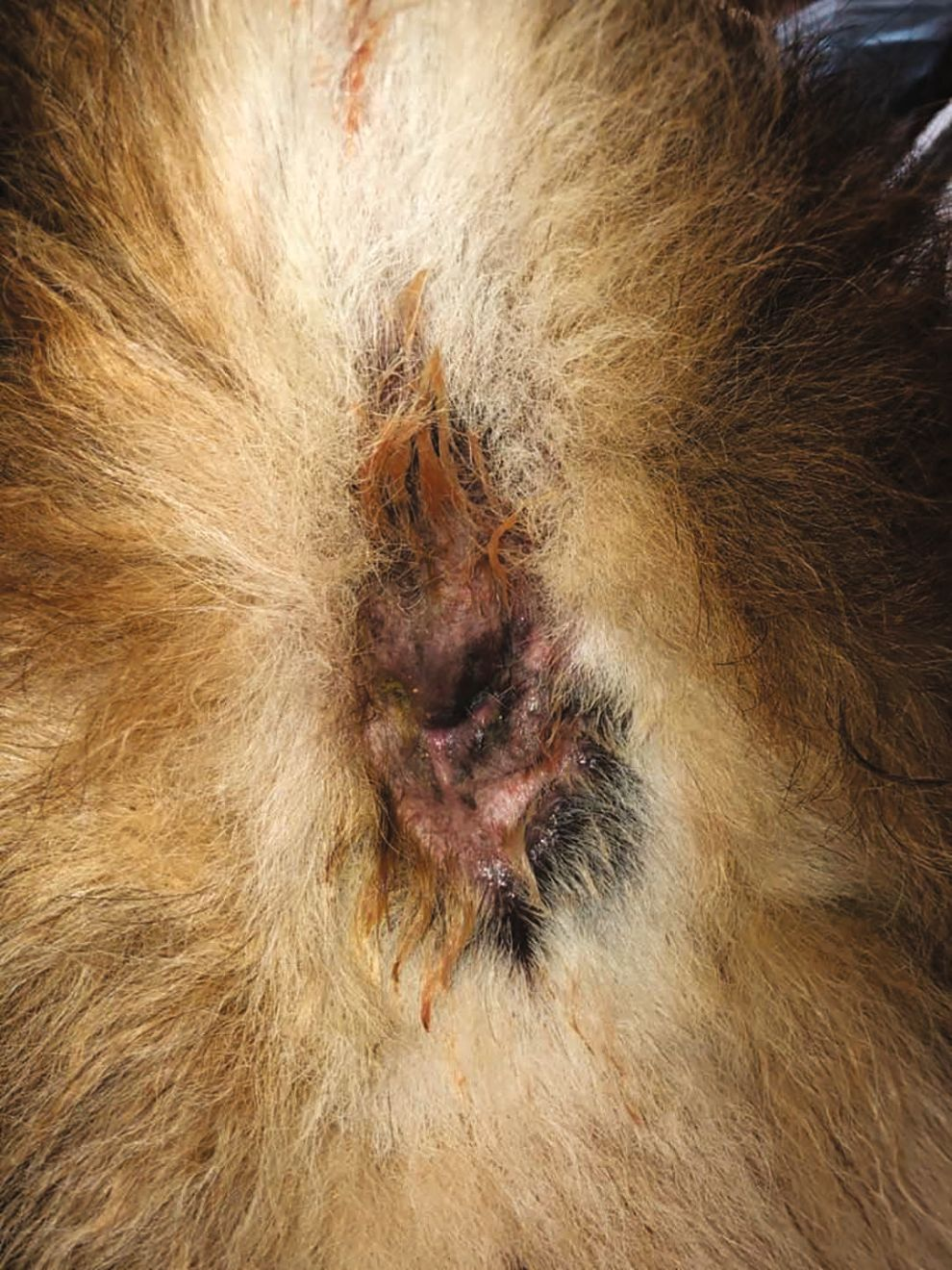
Another reported application of FBM is for canine perianal fistulae [16]. In this study, four dogs with the condition were treated solely with FBM once a week, with two consecutive applications at each session. After two weeks of treatment all dogs had improved, with a significant reduction in vocalization, straining, and licking, and the perianal lesions were significantly decreased after five weeks of therapy. Again, the mechanism as to how FBM helped in these cases remains unknown. Figure 5 shows a case of canine perianal fistula before treatment with a commercially available FBM system once weekly. Three weeks after the first treatment there was reduction in the erythema and improvement of the fistula (Figure 6). It is, therefore, speculated that the effects of FBM on wound healing might be beneficial in such cases [1],[2],[4].
It is important to mention that many of the above conditions result from underlying diseases, such as atopic dermatitis, cutaneous adverse food reactions, ectoparasites, endocrine disorders and conformational problems. It is essential that wherever possible the underlying disease should be identified with a proper work-up (skin scrapes, cytology, blood tests, elimination diet, etc.) and properly addressed.
Finally, FBM has been reported to aid healing of acute uncomplicated surgical wounds [4]; FBM therapy has been shown to stimulate the release of promoting wound-healing cytokines and improve the microscopic characteristics of incisional wounds. Tissue re-epithelization was complete, with better collage deposition and less dermal inflammation. However, the macroscopic appearance of the wounds was not affected by FBM [4] and further, larger studies are needed to evaluate these results [9].
FBM has been applied with promising results in treating both superficial bacterial folliculitis and deep pyoderma in dogs.
Conclusion
These promising findings suggest that FBM can be used to treat several canine dermatological disorders and that there is the potential to expand its applications in veterinary medicine. Further studies are, however, needed to evaluate the efficacy of the modality in treating different skin diseases and to reveal its exact mechanism of action.
References
- Marchegiani A, Spaterna A, Cerquetella M. Current applications and future perspectives of fluorescence light energy biomodulation in veterinary medicine. Vet. Sci. 2021;8:20. https://doi.org/10.3390/VETSCI8020020
- Scapagnini G, Marchegiani A, Rossi G, et al. Management of all three phases of wound healing through the induction of fluorescence biomodulation using fluorescence light energy. BiOS. 2019;31. https://doi.org/10.1117/12.2508066
- Hill PB, Lo A, Eden CAN, et al. Survey of the prevalence, diagnosis and treatment of dermatological conditions in small animals in general practice. Vet. Rec. 2006;158:533-539. https://doi.org/10.1136/VR.158.16.533
- Salvaggio A, Magi GE, Rossi G, et al. Effect of the topical Klox fluorescence biomodulation system on the healing of canine surgical wounds. Vet. Surg. 2020;49:719-727. https://doi.org/10.1111/VSU.13415
- Marchegiani A, Spaterna A, Fruganti A, et al. Exploring fluorescent light energy as management option for canine superficial bacterial folliculitis. Front. Vet. Sci. 2023;10:1155105. https://doi.org/10.3389/fvets.2023.1155105
- Marchegiani A, Fruganti A, Spaterna A, et al. The effectiveness of fluorescent light energy as adjunct therapy in canine deep pyoderma: A randomized clinical trial. Vet. Med. Int. 2021;6643416. https://doi.org/10.1155/2021/6643416
- Apostolopoulos N, Mayer U. Use of fluorescent light energy for the management of bacterial skin infection associated with canine calcinosis cutis lesions. Vet. Rec. Case Rep. 2020;8:e001285. https://doi.org/10.1136/VETRECCR-2020-001285
- Marchegiani A, Spaterna A, Cerquetella M, et al. Fluorescence biomodulation in the management of canine interdigital pyoderma cases: a prospective, single-blinded, randomized and controlled clinical study. Vet. Dermatol. 2019;30:371-e109. https://doi.org/10.1111/VDE.12785
- Perego R, Mazzeo M, Spada E, et al. Critically appraised topic on low-level laser therapy (LLLT) in dogs: An advisable treatment for skin diseases? Vet. Sci. 2022;9:505. https://doi.org/10.3390/VETSCI9090505
- Marchegiani A, Fruganti A, Gavazza A, et al. Fluorescence biomodulation for canine interdigital furunculosis: updates for once-weekly schedule. Front. Vet. Sci. 2020;9:880349. https://doi.org/10.3389/fvets.2022.880349
- Lundberg A, Hathcock T, Kennis RA, et al. In-vitro activity of fluorescence photobiomodulation therapy on meticillin-susceptible and -resistant Staphylococcus pseudintermedius and S. aureus; NAVDF congress abstracts. In: Vet. Dermatol. 2023;249-265.
- Schnedeker AH, Cole LK, Lorch G, et al. In-vitro bactericidal activity of blue light (465 nm) phototherapy on methicillin-susceptible and methicillin-resistant Staphylococcus pseudintermedius. Vet. Dermatol. 2017;28:463-e106. https://doi.org/10.1111/VDE.12451
- Tambella AM, Attili AR, Beribè F, et al. Management of otitis externa with a led-illuminated gel: A randomized controlled clinical trial in dogs. BMC Vet. Res. 2020;16:1-14. https://bmcvetres.biomedcentral.com/articles/10.1186/s12917-020-02311-9
- Saridomichelakis MN, Farmaki R, Leontides LS, et al. Aetiology of canine otitis externa: a retrospective study of 100 cases. Vet. Dermatol. 2007;18:341-347. https://doi.org/10.1111/J.1365-3164.2007.00619.X
- Gil N, Santoro D, Marsella R. In-vitro susceptibility of Malassezia pachydermatis to low-level light therapy; NAVDF congress abstracts. In: Veterinary Dermatology. John Wiley & Sons, Ltd, 2023;249-265
- Marchegiani A, Tambella AM, Fruganti A, et al. Management of canine perianal fistula with fluorescence light energy: preliminary findings. Vet. Dermatol. 2020;31:460-e122. https://doi.org/10.1111/VDE.12890

Neoklis Apostolopoulos
DVM, Dip. ECVD, University of Wisconsin-Madison, School of Veterinary Medicine, Madison, WI, USAUnited States
Dr. Apostolopoulos obtained his veterinary degree at the University of Thessaly in Greece. He then moved to Germany where he completed a rotating internship, a residency, and postgraduate research with a focus on canine cutaneous microbiota. He is currently an assistant clinical professor of veterinary dermatology at the University of Wisconsin-Madison, with a research focus on the canine skin microbiome and applied artificial intelligence in veterinary dermatology. His clinical interests include all aspects of dermatology, allergology, and otology, and he also has experience in CO2 laser surgery and fluorescence photobiomodulation treatment of skin diseases.