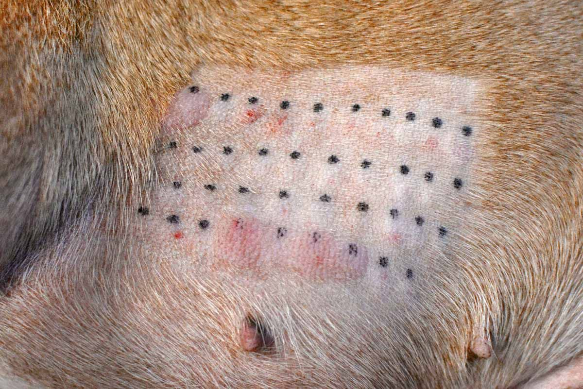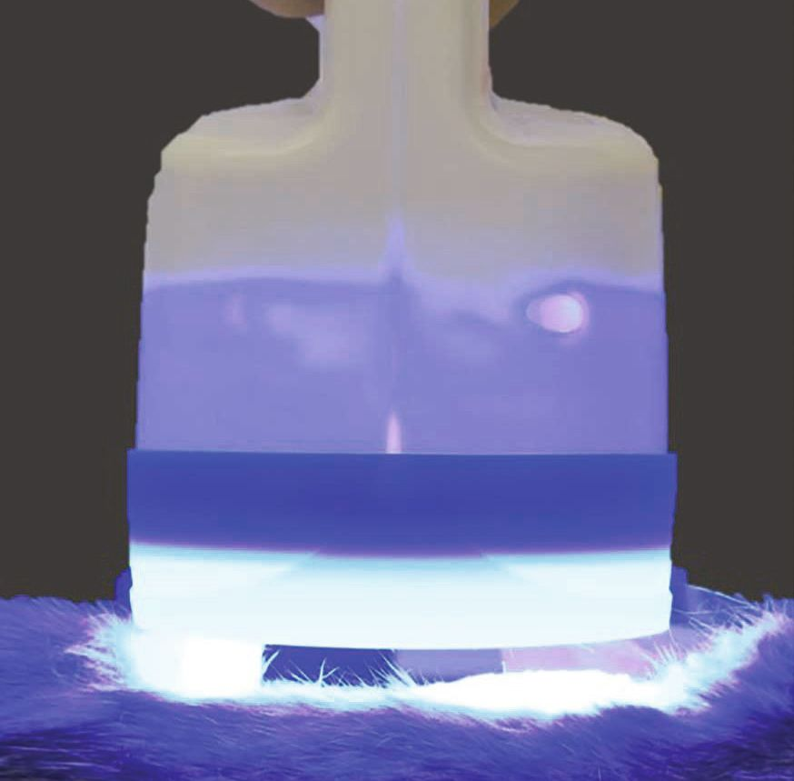
Introduction
Describing a clinical condition as “chronic” can mean that it has a prolonged time course, that definitive healing is impossible, or that the underlying cause is persistent. Any of these definitions can apply to otitis [1] when treatment has been ineffective due to insufficient control of the primary cause of the disorder, or because there is a perpetuating element that has not been detected or has not been adequately resolved [2]. This article will review why the canine ear is predisposed to otitis, and what can be done to minimize the risk of an ear problem becoming chronic.
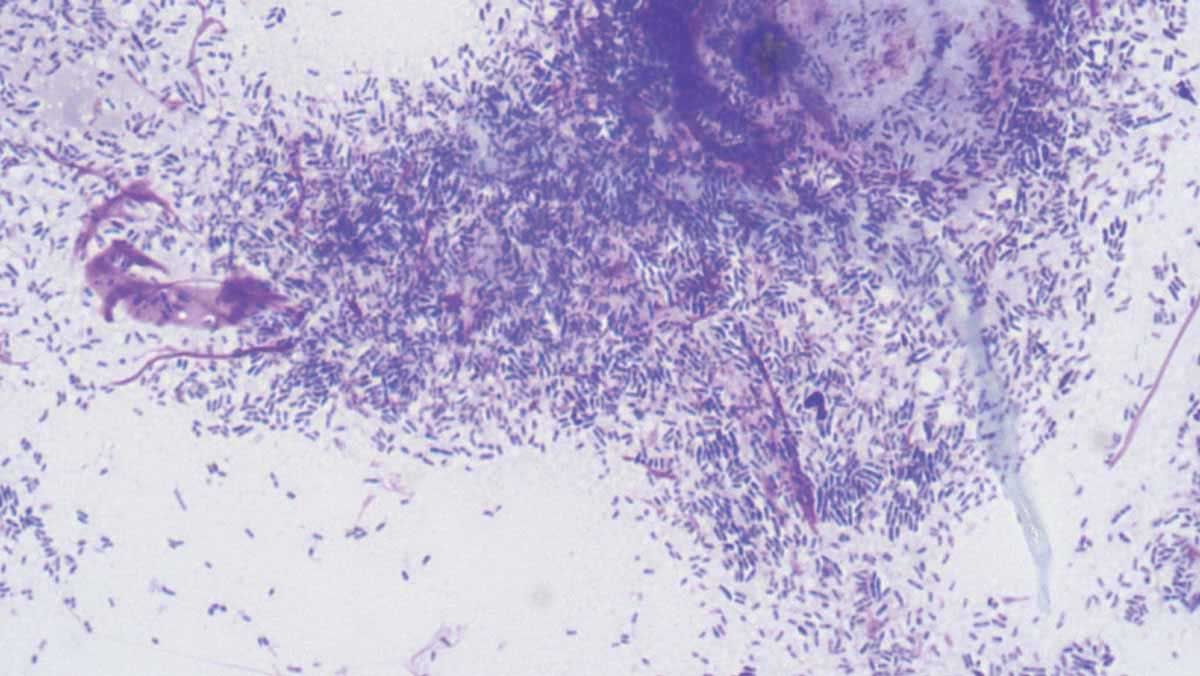
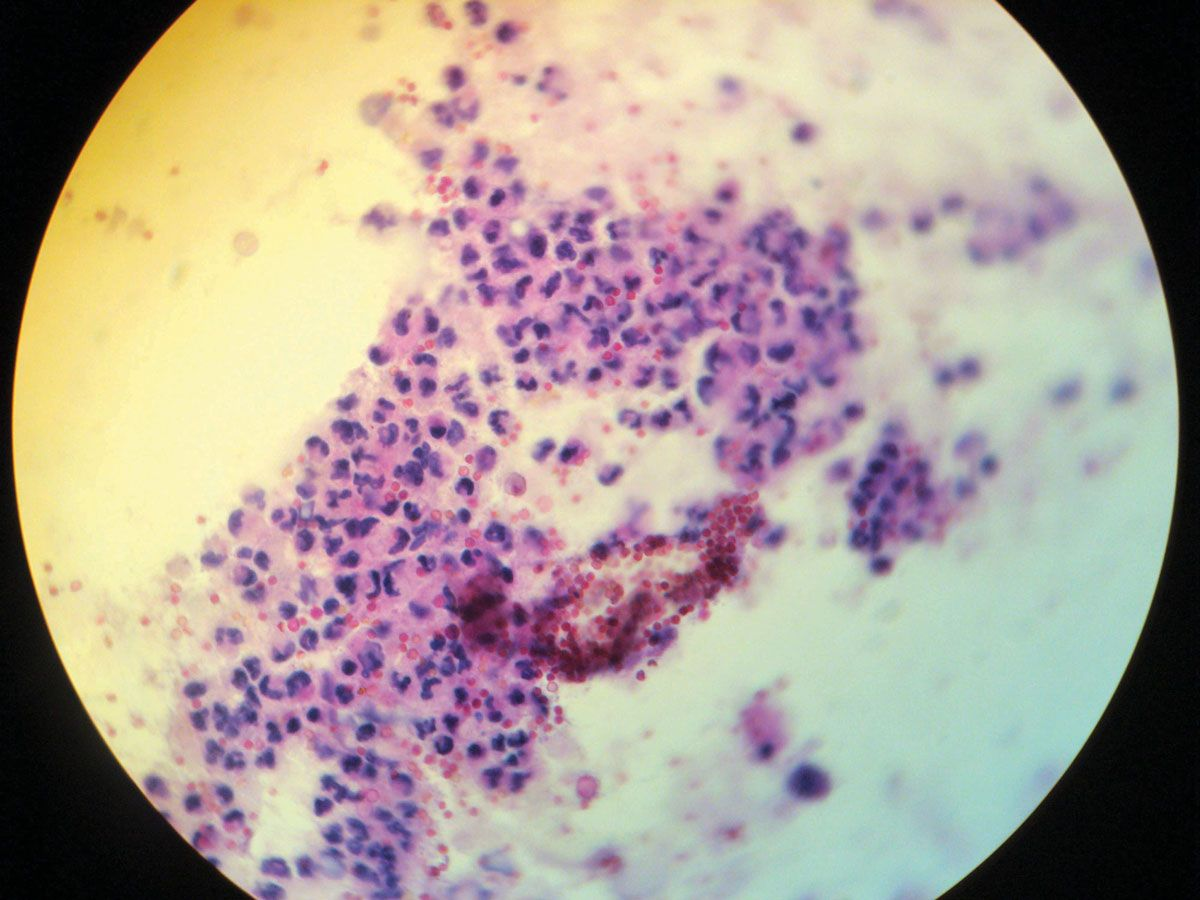
Firstly, it should be said that the anatomical structure of the ear in itself favors dysbiosis of the microbiome, as intertrigo (inflammation caused by the rubbing of one area of skin on another) problems can easily develop, and its “anti-gravitational” conformation discourages drainage [3],[4] (Figure 1). However, not all cases of otitis will necessarily lead to dysbiosis, and in some situations abundant cerumen production and itching can be taken to constitute otitis. In addition, secondary agents (Figures 2 and 3) will proliferate when the ear’s defense and microbiome controls fail, as can be seen from cytological studies [5],[6]; indeed, cytology must be undertaken for all cases of otitis in order to establish a firm diagnosis, and some tips are given in Table 1.
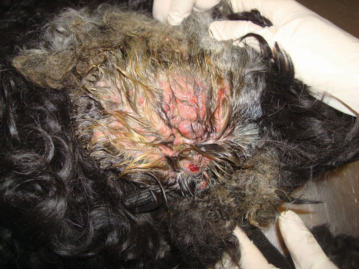
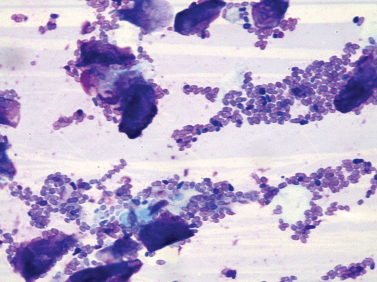
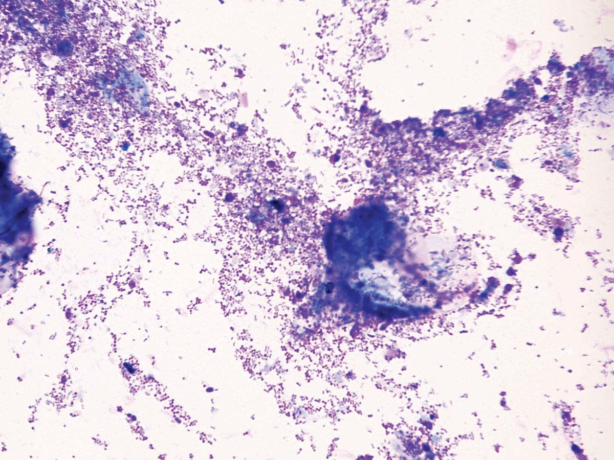
Table 1. Some tips for interpreting cytology from ear swabs.
|
What causes acute or chronic otitis?
The most common mistakes made by veterinarians, and which may lead to otitis becoming chronic, include:
- Suspending treatment based only on clinical improvement, without cytological confirmation.
- Using antibiotics at random without first obtaining an antibiogram.
- Failing to perform thorough cleaning despite adequate antibiotic treatment.
- Failing to proactively treat the primary cause of the otitis, thereby facilitating recurrence.
- Being limited in treatment options because of economic or logistical issues (i.e., cost or the prolonged nature of therapy).
It is, therefore, very important to remember the causes that underlie altered ear homeostasis which can give rise to acute inflammation; if not effectively controlled, these can result in structural changes to the external canal and/or middle ear, and perpetuation of the problem [2]. All studies on canine otitis mention certain factors inherent to affected patients and which predispose to the condition. Such characteristics are commonly found in the canine population and will favor the development of otitis [7]; they include (from greatest frequency):
- Atopic dermatitis
- Foreign bodies (grass fragments or ceruminous otoliths)
- Adverse food reactions
- Infestation due to Otodectes cynotis (ear mites)
- Polyps and tumors of the external auditory canal
- Adverse reactions to topical ear medications
- Endocrine disease
- Primary seborrheic disorders
- Transient ecosystem changes (due to inadequate care)
- Aural demodicosis (or other less frequent parasites)
- Aural dermatophytosis
Apart from these primary causes, the presence of predisposing factors may contribute to chronicity of the problem, since they favor recurrence and complicate treatment, causing the latter to be less effective. These include:
- Frequent swimming
- Conformational conditions such as a narrow, long, deep and/or hairy auditory canal
- Excessive cerumen production due to idiopathic causes
- Drooping external ears/pinnae
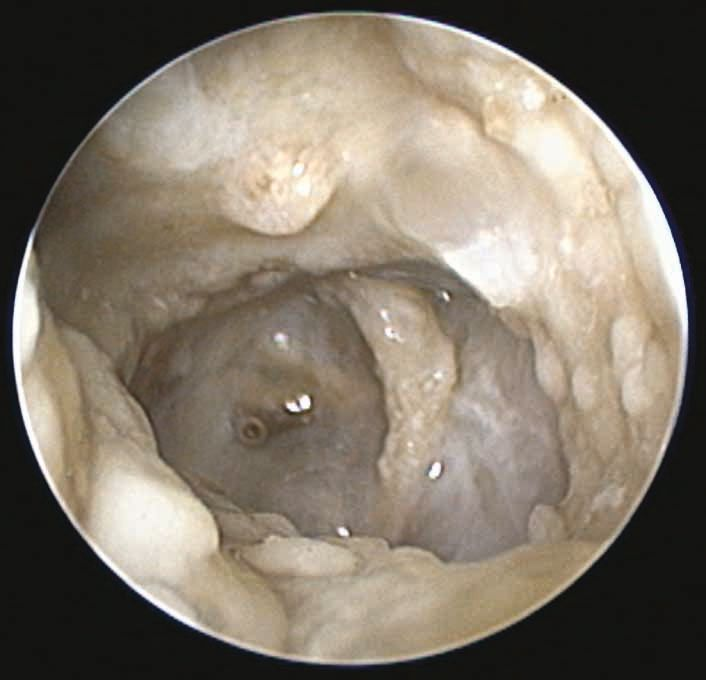
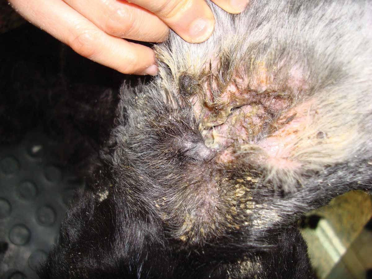
When otitis appears, the microenvironment conditions within the ear change, due to an increase in cerumen production and narrowing of the canal induced by the inflammatory process. This inevitably leads to alteration of epithelial migration, resulting in dysbiosis of the microbiome. If this is not quickly resolved, the changes may persist, causing the otitis to become chronic (Figure 4).
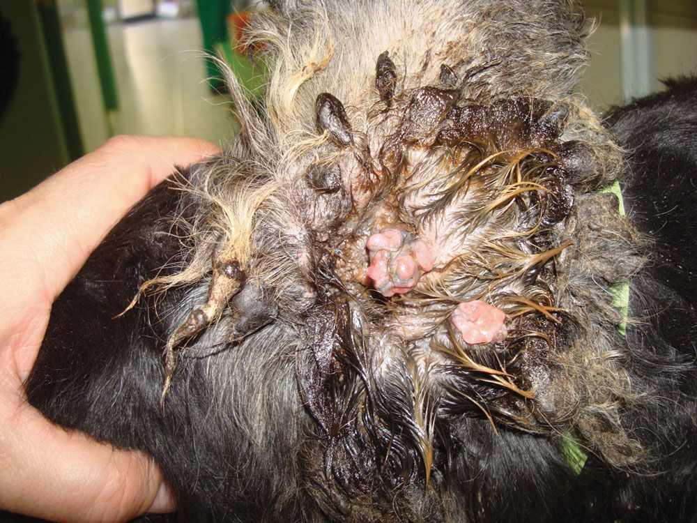
Most cases of otitis start with the appearance of aforementioned primary factors, and failure to resolve the problem may lead to persistence due to various conditions developing subsequently [8]. These can include:
- The favoring of resistant bacterial strains due to incorrect treatment.
- Eardrum rupture from enzymes secreted by invasive bacteria.
- Perpetuation of the infection due to unresolved otitis media.
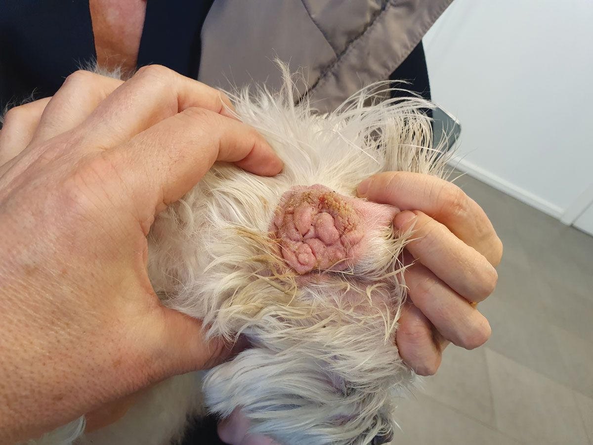
- Hyperplasia, fibrosis, narrowing and calcification of the auditory canal over time (Figure 5).
- The accumulation of cerumen (otoliths) that obstruct epithelial migration and sequestrate bacteria.
- The organization of microorganisms into treatment-resistant biofilms.
- Excessive or irritating ear cleaning, which may favor inflammation.
Preventing acute otitis from becoming chronic
Primary causes
If the primary cause can be identified, this will aid in selecting the most appropriate treatment.
- Atopic dermatitis: This disease clearly induces an imbalance in the ear’s microenvironment. The cerumen of affected dogs may fail to control the microbiome, not only due to its excess production secondary to inflammation, but also because of a deficiency in defensins (host defense peptides). Avoiding relapse in the form of outbreaks that can disrupt the microenvironment is crucial in order to prevent ceruminous otitis with or without dysbiosis from repeating. Each individual requires a personalized strategy, but proactive measures with systemic medication are recommended, for example monthly lokivetmab injections, daily oclacitinib or cyclosporine at maintenance doses, corticosteroids at pulse dosing (e.g., twice weekly), or anti-fungal azoles (again pulse dosing). Topical treatment may also be appropriate for some cases; these include corticosteroid preparations, antiseptic agents, anti-fungal azoles, or cerumenolytic agents to keep the canal clear, all administered once or twice a week. Topical corticosteroids can control inflammation and the toxic negative effects of excess cerumen, allowing the microbiome to exert its protective function correctly; in many cases dysbiosis can be controlled without the use of antiseptics or antibiotics.
- Foreign bodies: Clinical experience shows that there are cases of otitis in which a foreign body or an otolith has remained lodged for a long period of time, and is only discovered when performing otoscopy under sedation or general anesthesia. A thorough evaluation of the ear is, therefore, highly recommended in order to rule out any possible obstruction of the canal or to detect the presence of excessive post-inflammatory cerumen.
- Adverse food reactions: Failure to identify a dietary allergen may give rise to or perpetuate otitis, causing it to become chronic. When following a diagnostic protocol with a relapsing otitis, this possibility should be examined via the use of an elimination diet.
- Ear mites: In recent years the introduction of macrocyclic lactones and isoxazolines as routine external antiparasitic agents has caused otoacariasis to become infrequent. However, the diagnosis is relatively simple and is based mainly on the type of cerumen produced.
- Polyps and tumors: Polyps develop mainly as a consequence of chronic irritation, whilst various different tumors can be found in the ear canal. Any growth will hinder epithelial migration and favor chronic dysbiosis. When they involve the middle ear, these lesions may be more difficult to diagnose, and can contribute to ongoing inflammation or chronic infection in the area.
- Adverse reactions to topical products: Worsening of otitis signs following topical treatment may be indicative of an adverse contact reaction to the drug employed. These will typically appear shortly after application of the product, with severe erythema and exudation developing, mainly at the entrance to the canal.
- Endocrine disease: hormone abnormalities, especially hypothyroidism but also hyperadrenocorticism or altered sex hormones, can adversely affect cerumen secretion and favor dysbiosis, with the subsequent development of varying degrees of otitis.
- Primary seborrheic disorders: These are more common in some breeds of dog and can alter the ear ecosystem due to changes in the quality and amount of cerumen (Figure 6).
- Transient ecosystem changes: In patients that are susceptible to otitis due to the presence of one or more primary causes, inappropriate cleaning or aesthetic care may serve as a trigger to induce or perpetuate otitis. For example, plucking intra-auricular hairs, excessive or very irritating cerumenolytic use, or the use of swabs can all facilitate ecosystem imbalance [8].
- Otic demodicosis: Nowadays this condition is rare, and as with Otodectes, newer antiparasitic medications have caused it to become even more unusual. However, the presence of Demodex mites in the outer ear alone may be considered, and will always be a possibility in dogs that have been treated for parasites with substances other than macrocyclic lactones or isoxazolines.
Predisposing factors
When it comes to predisposing factors, certain measures can be taken to prevent otitis from becoming chronic [9]. For instance:
- Frequent swimming: Avoiding this habit altogether may be a good option; however, if it proves difficult, the use of topical antiseptics or pH acidifiers applied to the ears when the dog comes out of the water may help counter the development of dysbiosis.
- Conformational conditions: Certain breeds have anatomical characteristics that predispose to otitis; the Shar Pei is most commonly implicated. Clinical experience has shown that if no intervention is necessary, it is best not to use cleaning agents in their ears. Where a primary anatomical cause induces acute inflammation, keeping the ear canal clear can be difficult, and in many situations it will be necessary to apply topical corticosteroids and cerumenolytic agents on a regular basis.
- Hairy auditory canals: Again, this is often a breed-specific problem – e.g., in the Bichon Frise. Otoscopy will show hair extending to the periphery of the tympanic membrane. In these dogs, although keeping the canal clear may require removal of the hair, where a primary cause is present, it may be better to avoid this practice in order to avoid follicular microtrauma.
- Narrow, long or deep auditory canals: These features are one of the most important causes contributing to the complication of otitis, and again can be breed-specific (e.g., the German Shepherd dog); for example, a long canal increases detritus accumulation and makes it more difficult to remove.
- Excessive cerumen production: In some breeds or individuals, excessive cerumen may be directly related to generalized seborrhea, and it is not always easy to determine the cause. These patients will need regular application of cerumenolytic agents and topical corticosteroids to reduce the amount of cerumen produced.
- Drooping external ears: Pinnae that cover access to the ear canal can complicate all treatments and prevent moist and detritus-laden ears from aerating and releasing these products.
Gustavo Machicote Goth
Perpetuating factors
The above recommendations can serve both to avoid otitis and to prevent it from repeating or becoming chronic. However, it is important to take into account that recurrence sometimes cannot be avoided due to the existence of perpetuating factors. Three main ones have been identified: biofilm formation, otitis media, and narrowing, fibrosis and calcification of the ear canal.
- Biofilm formation
Both yeasts and some bacterial strains (mainly Pseudomonas spp.) can organize into a layered formation along with glycoproteins and air and fluid distribution channels that are resistant to traditional cleaning practices and antibiotics, thereby allowing perpetuation (Figure 7). It is known that the bacterium Finegoldia magna in the external auditory canal is an opportunistic anerobic pathogen that facilitates Pseudomonas biofilm formation [10]. It is sometimes not easy to identify biofilm, although there are some features suggestive of its presence, such as a dark, sticky, and bright mucoid secretion that does not adequately respond to treatment. Although biofilm can be reduced with treatment, microorganism overgrowth is never fully controlled.
Acetylcysteine and Tris-EDTA may serve to disrupt the biofilm, mainly by acting on the sulfur bonds that stabilize it. In the presence of an intact eardrum, liquids with potent cleaning action, such as carbamide peroxide, can also be used. According to some studies, enzymatic cleaning agents such as lactoferrin, lactoperoxidase and lysozyme may be effective in cases of otitis with resistant strains. New Burow’s solution (based on 2% aluminum acetate and 0.1% betamethasone) may be indicated in cases of otitis media with infection due to multi-resistant strains [11]. Another option is video-otoscopy under general anesthesia to perform circular brushing using a pump and saline suction [12].

- Otitis media
Middle-ear problems are a common complication of chronic otitis, particularly when gram-negative bacteria such as Pseudomonas spp. are present [13]. The proteolytic enzymes released by these bacteria can rupture the eardrum, spreading the infection and the inflammatory products into the middle ear. A biofilm may also develop at this site, sometimes affecting the tympanic bulla and generating a situation that considerably complicates the prognosis [1],[14]. Cytological study is fundamental in all patients with otitis media, and differentiation between cocci and bacilli can greatly guide the steps to be taken [15],[16]. It is very important to take into account that in allergic dogs, Malassezia overgrowth may perpetuate chronicity, and in some cases can prove very difficult to treat, partly again as biofilm formation may be involved [1],[14],[15],[16]. In addition, although culture and an antibiogram are essential in these circumstances, sometimes there is no direct relationship between the in-vitro and in-vivo findings of these tests [1],[5],[9],[10]. Experience shows quinolones to be the safest antibiotics to use where there is a ruptured tympanic membrane, and they usually exhibit a broad spectrum of action. The use in the mornings of a combination of cleansing fluid composed of Tris-EDTA and/or n-acetylcysteine is usually the preferred method for eliminating inflammatory debris, rupturing the biofilm and sensitizing the bacteria to the antibiotics that will be administered afterwards.
Suspicion of otitis media with a ruptured tympanic membrane may be raised by a number of signs:
- Coughing or swallowing at the time of washing.
- Pain on pressing the throat near the bulla.
- The formation of bubbles when washing and flooding the external ear.
- Deeper penetration of a probe when comparing the healthy ear to the ear with the ruptured eardrum.
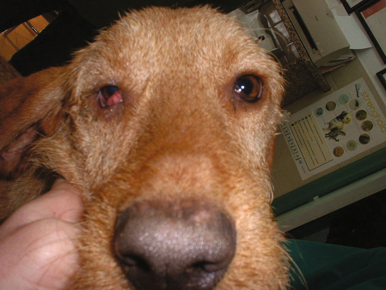
- Presence of neurological signs (Figure 8).
- Otitis unresponsive to treatment and which shows some opacification of the lumen of the bulla or wall thickening on radiography.
However, the best diagnostic images can be obtained with magnetic resonance imaging or computed tomography [17].
- Alterations to the ear canal
Chronic inflammation of the ear canal, including the dermis and the cartilage of the external ear, causes the tissues to fibrose, with the accumulation of calcium salts that result in a rigid and irreversible structure. In the early stages, or before the calcium accumulates, when narrowing is due mainly to edema of the canal’s subdermal layer, systemic corticosteroids at immunosuppressive doses for 15 days may be used in an attempt to reverse the stenosis and allow restoration of epithelial migration [14]. In addition, 0.1% mometasone cream or lotion applied topically may help open the ear canal.
Managing patients prone to relapse
There are situations in which the balance of the ear’s ecosystem is very sensitive to relapse, or the structural changes are so serious that restoration of epithelial migration is impossible and the options for avoiding ablation surgery are minimal. In some circumstances pulse-based therapy is inevitable, such as where:
- Cytology persistently shows microorganism overgrowth with abundant cerumen and keratinocytes.
- The ear canal lumen remains narrow, and cleaning cannot be easily achieved.
- The tympanic membrane fails to heal, and cytology of the middle ear continues to show the presence of microorganisms, even if no inflammatory cells are observed.
Pulse-based treatments usually involve effective cerumenolytic agents combined with antifungal drugs and antiseptics to control overgrowth and prevent proliferation. In some cases, corticosteroids (e.g., hydrocortisone aceponate) can help, and may be a good way to avoid excessive cerumen production and secure reduced levels of inflammation [18]. Conditions that prove difficult to treat may arise in some patients with chronic otitis overtreated with antibiotics and which ultimately suffer invasion by fungal organisms such as Aspergillus spp. These cases are characterized by ulceration and other severe signs, and require thorough cleaning and intensive therapy with antifungals such as azoles via both topical and systemic routes [14] (Figures 9 and 10).
In other cases, healing is impossible because of terminal situations such as perpetual biofilms that do not respond to treatment (Figures 11-13) or where stenosis of the ear canal is so severe that anti-inflammatory medication proves ineffective. When the restoration of epithelial migration is not possible due to irreversible structural changes, there may be no option other than surgical exposure of the ear canal and ablation of the bulla with curettage (Figure 14) in order to avoid relapses (Table 2). However, surgical measures are not always a definitive solution, since in some cases the primary causes continue to act on small areas of epithelium of the residual canal. Owners must understand that surgery should always be the last option, and the clinician should be aware that this measure may be interpreted as a failure of the previous prescribed medical treatment.
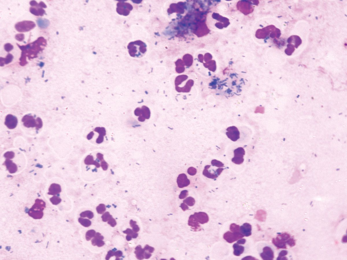
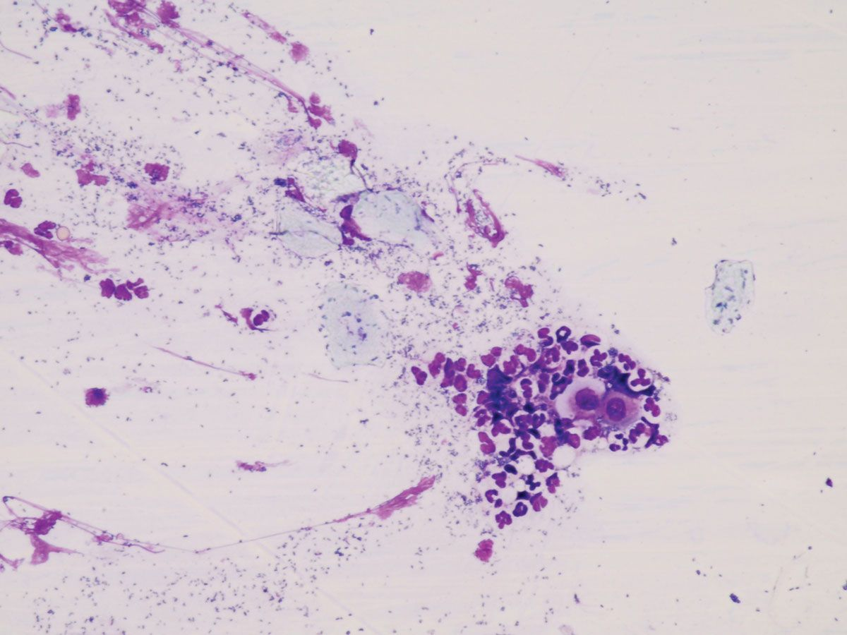
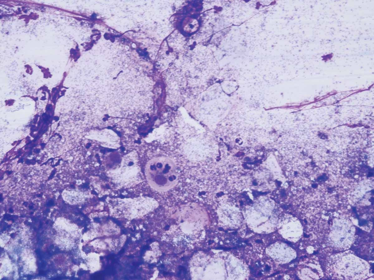
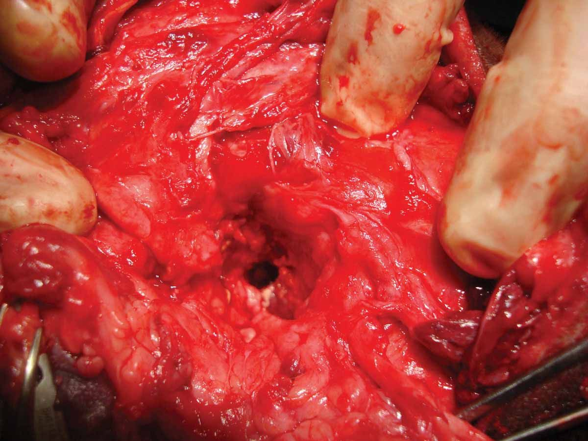
Table 2. Surgical options for chronic otitis externa ([1],[14],[19],[20]).
|
Lateral wall resection, for:
|
|
Total ablation of the ear canal and osteotomy of the bulla, for:
|
Conclusion
Prevention is always better than cure, and nowhere more so than with otitis. The clinician should be aware of the primary causes of otitis and be able to identify dogs at risk due to the presence of predisposing factors. Appropriate and aggressive treatment, linked to careful cytological testing, should help minimize the chances of an acute otitis becoming chronic and possibly irreversible.
References
- Hill, P. Management of chronic infection. In; Proceedings, WCVD Congress. Sydney 2020.
- Apostolopoulos, N. The Canine Skin and Ear Bacterial Microbiota. Todays Vet Pract. May/June 2023.
- Zalke A. Clinical efficacy of Neo Burows solution in six dogs with suppurative otitis media. In; Proceedings, WCVD Congress Sidney 2020.
- Noxon J. Canine Otitis Externa Best Clinical Practices: Clinical Consensus Guidelines of the World Association for Veterinary Dermatology. In; Proceedings, ESVD Congress Porto 2021.
- Shell LG. Otitis media and otitis interna: Etiology, diagnosis, and medical management. Vet. Clin. North Am. Small Anim. Pract. 1988;18(4):885-899.
- Miller Jr. WH, Griffin CE, Campbell KL. Miscellaneous skin diseases. In Muller and Kirks Small Animal Dermatology 7th ed. Missouri: Elsevier Mosby, 2013;708-709.
- Lipscomb H, de Bellis F. A diagnostic approach to canine otitis. Vet. Focus 2021;32(1).
- Angus JC. Otic cytology in health and disease. Vet. Clin. North Am. Small Anim. Pract. 2004;34:411-424.
- Sturges BK, Dickinson PJ, Kortz GD, et al. Clinical signs, magnetic resonance imaging features and outcome after surgical and medical treatment of otogenic intracranial infection in 11 cats and 4 dogs. J. Vet. Intern. Med. 2006;20:648-656.
- Niza Virbac Otology Meeting 2023.
- Lanz OI, Wood BC. Surgery of the ear and pinna. Vet. Clin. Small Anim. Pract. 2004;34(2):567-599.
- Griffin CE. Otitis techniques to improve practice. Clin. Tech. Small Anim. Pract. 2006;21(3):96-105.
- Radlinsky MG, Masan DE. Enfermedades del odo. In; Tratado de medicina interna veterinaria. Enfermedades del perro y el gato. Ettinger SJ, Feldman EC (eds). Madrid; Elsevier Espaa S.A.;2007;1168-1186.
- Getty R. Anatomy of the canine. In: Sisson and Grossmans Anatomy of Domestic Animals. Philadelphia, WB Saunders. 1996;1214-1245.
- Blauch B, Strafuss AC. Histologic relationship of the facial (7th) and vestibulocochlear (8th) cranial nerves within the petrous temporal bone in the dog. Am. J. Vet. Res. 1974;35:481.
- Ngo J. Taminiau B. Fall PA, et al. Ear canal microbiota a comparison between healthy dogs and atopic dogs without clinical signs of otitis externa. Vet. Dermatol. 2018;29:425-e140.
- Boynosky NA, Stokking LB. Retrospective evaluation of canine dermatitis secondary to Corynebacterium spp. J. Am. Anim. Hosp. Assoc. 2015;51(6):372-379.
- Gotthelf NL. Enfermedades del odo. In; Animales de Compaa. Buenos Aires; Intermdica. 2001;26-32.
- Carlotti DN. Diagnosis and medical treatment of otitis externa in dogs and cats. J. Small Anim. Pract. 1991;32(8):394-400.
- Bajwa J. Canine otitis externa: treatment and complications. Can. Vet. J. 2019;60(1):97-99.
Gustavo Machicote Goth
DVM, MS (oncology), Clínica Veterinaria Vilanova, Pontevedra, Spain
Dr. Machicote Goth qualified from the University of Buenos Aires and is currently employed at clinics in Pontevedra and Santiago de Compostela with responsibility for dermatology. He is accredited in dermatology by AVEPA (Asociación de Veterinarios Españoles Especialistas en Pequeños Animales) and is a member of the Scientific Committee of GEDA (the AVEPA Dermatology Group), serving as secretary between 2004-2008. He has led various training courses in both Europe and America, and has published numerous articles on dermatology, as well as co-authoring several textbooks.