Introduction
Canine atopic dermatitis (CAD) is a common inflammatory skin disease, affecting up to 15% of the global dog population [1]. The pathogenesis of the disease is multifactorial, with both skin barrier dysfunction and immunological dysregulation known to have central roles, and both may be influenced by genetic and environmental factors. IgE and non-IgE mediated immunological events are key features in the pathogenesis, with allergens constituting the main triggering factors [2]. The most commonly associated laboratory feature in CAD is the allergen-specific serum IgE levels, but (in contrast to humans) elevated total IgE levels do not assist in the diagnosis of CAD. Dogs are reported to have much higher levels of IgE than humans, probably as a result of their more frequent exposure to parasite infestation [3].There are two major risk factors for atopic dermatitis; breed predisposition (e.g., 50% of West Highland White terriers may be affected) and a familial history of CAD [4]. However, since both genetic and environmental factors are involved, the phenotypic manifestation of the disease is highly variable – not only between different breeds, but also among individual dogs of the same breed. Given that CAD is both a complex disease with multiple facets and that other skin conditions may mimic the condition, a definitive clinical diagnosis is considered challenging.
Diagnostic considerations
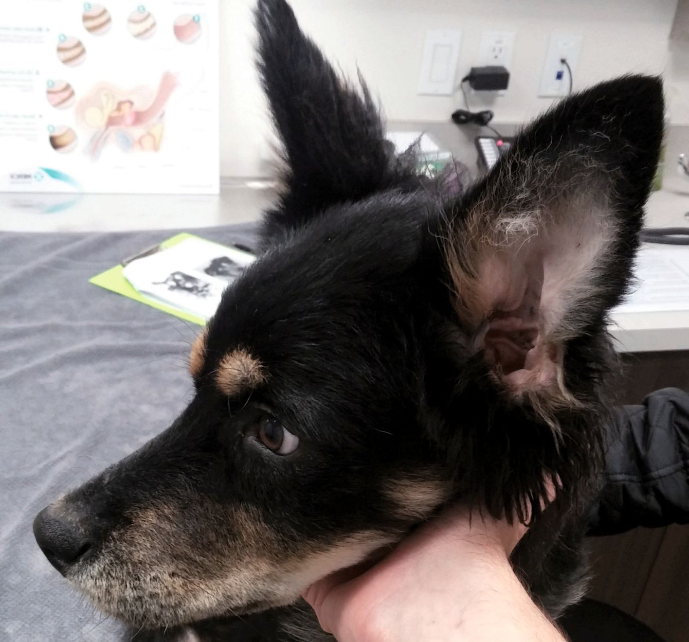
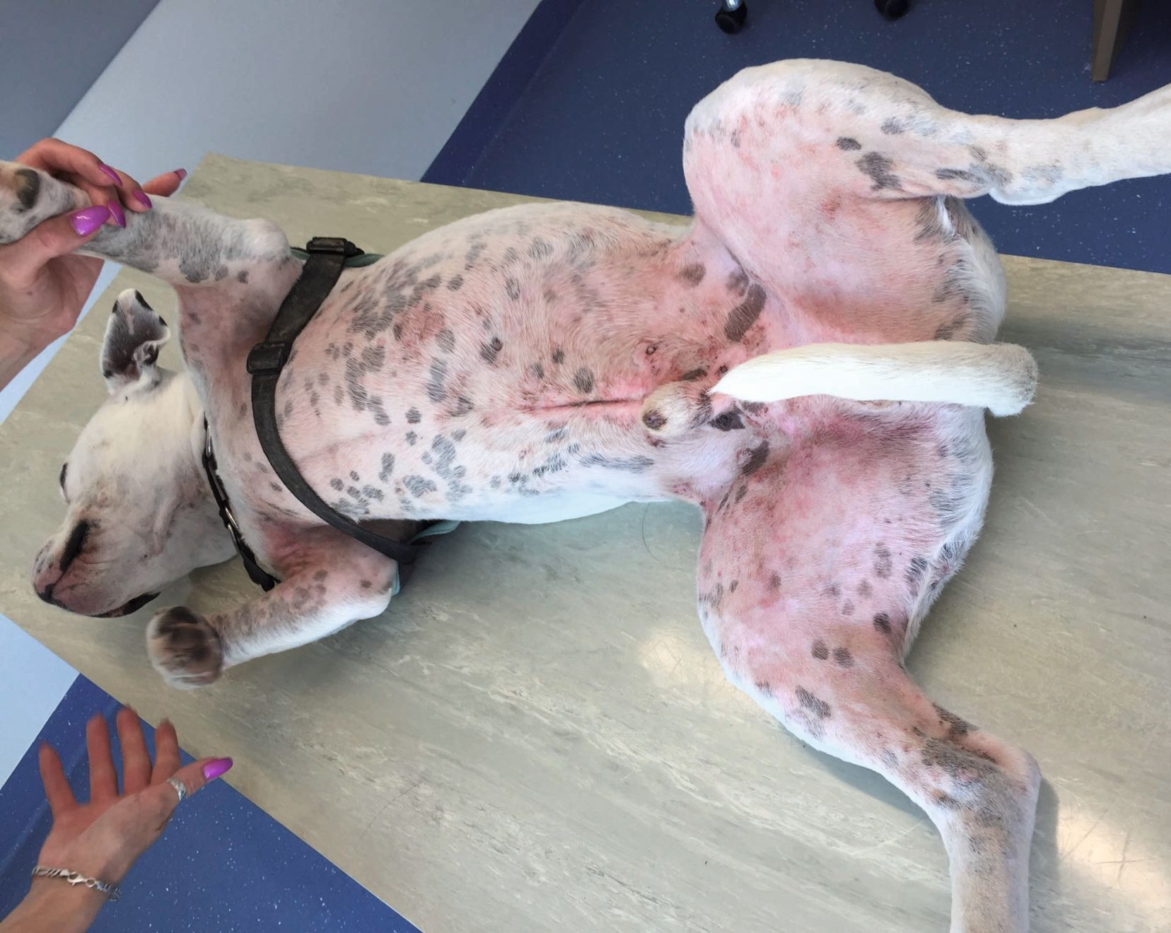
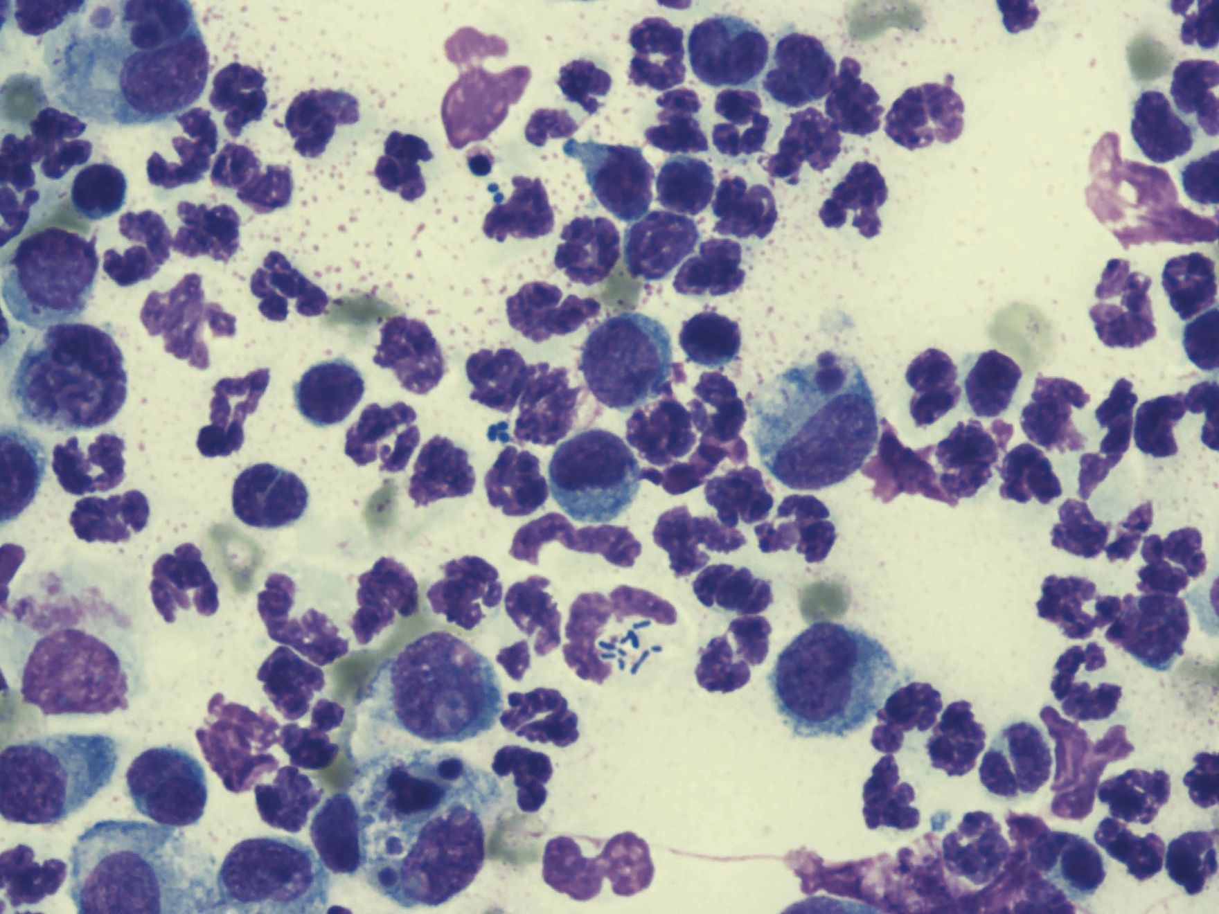
Because there is currently no reliable biomarker that can distinguish CAD from other dermatological disorders, the diagnosis of CAD remains clinical, and hence the clinician must interpret and consider various aspects, including the patient’s history, characteristic clinical features and exclusion of other differential dermatoses. Figure 1 offers a workflow for the diagnosis of CAD. The first step is to rule out other CAD-mimicking diseases, because although pruritus is the most consistent finding, it is by no means exclusive for CAD, and other differentials should be considered. Ectoparasite infestations or bacterial or yeast infections, secondary to a non-pruritic disorder (e.g., endocrinopathies, sebaceous adenitis), or less frequently neoplastic disease (e.g., cutaneous lymphoma, though more commonly seen in older patients), should be ruled out during the initial workup phase on the basis of the signalment, history or additional targeted tests (Table 1). It is worth noting that one aspect very typical for CAD may be observed at the onset, when pruritus may be alesional or associated with primary skin lesions such as erythema and sometimes papules. With progression over time and additional secondary infections, signs such as pustules, alopecia, excoriations, lichenification, crusting and seborrhea may develop. The face, inner aspect of the pinnae, axillae, abdominal, inguinal and/or perineal areas and distal extremities are typical predilection sites in most dogs with CAD (Figure 2), although the affected body areas may vary with breed [5].
| Flea combing | Fleas |
| Skin cytology |
Malassezia dermatitis
Bacterial dermatitis
|
|
Skin scrapes/
hair plucking/
tape stripping
|
Scabies
Other ectoparasites: Demodex spp., Cheyletiella spp., Neotrombicula autumnalis
Dermatophytosis
|
| Fungal culture | Dermatophytosis |
| Skin biopsy |
Sebaceous adenitis
Cutaneous lymphoma
|
Once other potential etiologies have been ruled out, the standardized clinical criteria for CAD (“Favrot’s criteria”) can be applied to aid interpretation of the clinical findings in a pruritic dog (Table 2). These should not be employed before this point, because whilst ~80% of dogs that fulfil five of these criteria will have CAD, the remaining 20% will have another disease. Conversely, around 20% of dogs that do have CAD will not demonstrate at least five of these factors.
| Favrot’s criteria – the 8 major indicators for CAD [5] |
|---|
| History |
|
| Clinical exam |
|
| Specific clinical criteria for CAD |
|
Additional body sites which might be affected:
Recurrent skin/ear infections
|
Testing for environmental allergens
Once a clinical diagnosis of CAD has been made, further assessment is indicated, particularly to determine which allergens exacerbate clinical signs. This approach enables both appropriate selection of avoidance measures (especially with food allergens, although some measures can also be taken against house dust mites) and selection of allergens for allergen-specific immunotherapy. In general, if a dog has seasonal CAD, an immediate work-up for environmental allergens is warranted, but for cases with perennial pruritus and/or gastrointestinal clinical signs, food-induced dermatitis should be excluded before testing for environmental causes. An approach often used by the author is to initiate feeding of a commercial hydrolyzed diet using an elimination diet protocol. If the clinical signs of CAD persist despite this, testing for environmental allergens is followed, either by in vivo skin testing (most commonly intradermal testing, or IDT) or in vitro allergen-specific IgE serology (ASIS). Other than a poor response to a dietary trial, factors that would prompt allergy testing would be if a dog has severe clinical signs, where signs persist for more than 3 months each year, or if management with symptomatic therapy is unsuccessful (either because of significant drug side effects or poor owner compliance) [6].
It must be stressed that neither IDT or ASIS is a screening test for CAD; they only assist in confirming the clinical diagnosis and identification of allergens. Most dogs with CAD will have allergen-specific IgE to environmental allergens identified on testing, although in some cases IgE levels are not elevated (“atopic-like dermatitis”).
Both tests have their limitations and advantages, with neither being superior, and since the success rate of allergen-specific immunotherapy (ASIT) suggests that the two methods deliver comparable results [7] they may therefore be regarded as complementary. The author therefore prefers performing both skin testing and ASIS if costs allow, although if the former presents potential risks, or the patient is uncooperative, ASIS should be the initial option. If the two methods produce inconclusive results, the results are combined for ASIT, otherwise the choices for ASIT are generally based on the ASIS results. Importantly, for either method clinically relevant allergens must be chosen, which is very much dependent on the patient history and clinician’s judgement.
In addition, skin prick testing has recently become fashionable again, although as yet it is unvalidated in veterinary medicine. Saliva testing is also becoming commercially available, but at the time of writing it cannot be recommended as a diagnostic tool.
Intradermal testing (IDT)
IDT is an indirect measure of cutaneous mast cell reactivity, based on the presence of allergen-specific IgEs on their surface, and is the preferred diagnostic method among dermatologists, partly because mast cells can bind individual allergen-specific IgE molecules for more than a year [8]. Data on the sensitivity and specificity of IDT is scarce, although literature reports suggest it to be 30-90% and > 50-95% respectively [6] [9]. However, a precise assessment is very difficult due to a large number of both intrinsic factors (e.g., patient immunologic make-up) and extrinsic factors (e.g., allergen quality, skill level in performing IDT, season, medications).
Allergen selection
The selection of the most relevant allergens to be used for IDT depends on the animal’s geographic location, and may be aided by resources such as specialized veterinary and human clinics, allergy laboratories, textbooks and the relevant national allergy bureau. Nevertheless, the choice should be reviewed periodically, with individual allergens removed or incorporated as appropriate. For example, the author’s initial IDT panel, consisting of 43 allergens, has been reduced to the most frequently found 13 environmental allergens (Box 1), and is aligned with allergens used in the local human dermatology clinic. This revised panel has shown no reduction in the efficacy of ASIT over a seven-year period.
Grasses: Phleum pratense, Dactylis glomerata, Secale cereale
Trees: Fraxinus spp., Betula spp.
Weeds: Rumex crispus, Chenopodium album, Plantago lanceolata, Ambrosia spp., Artemisia vulgaris
|
IDT can utilize either lyophilized allergens or pre-diluted aqueous allergens intended for immunotherapy (which usually have a shelf life of at least 6-12 months), with the allergens further diluted as indicated in Table 3. They remain stable for up to 2 weeks if stored at 4°C in plastic syringes, or 8 weeks in glass vials, but otherwise allergen extract potency deteriorates with time [9], dilution and higher temperatures. Glycerinated allergens (usually used for prick tests in humans) should be avoided due to the possible irritative effects of the glycerin preservative.
| Allergens | Published concentrations/dilutions |
| Pollens | 1000 to 8000 PNU**/mL |
| Molds | 1000 to 8000 PNU/mL |
| House dust mites: |
|
|
D. pteronyssinus
|
100–200 PNU/mL
|
|
D. farinae
Tyrophagus putrescentiae
Lepidoglyphus destructor
|
75 PNU/mL
|
|
Acarus siro
Blomia tropicalis
|
50 PNU/mL
|
| Epidermal extracts |
At least 1,250 PNU/mL
300 PNU/mL for human dander
|
| Whole flea extract |
1:500 w/v |
Methodology
The only currently available recommendation for the optimal timing for IDT in dogs with seasonal disease is to test at the end or within 2 months of the peak season [10]; this avoids possible peak season anergy or out-of-season low IgE levels, although some dogs may show sufficient IDT reaction if tested during their peak season. Dogs with non-seasonal disease may be tested at any time of the year.
IDT can be performed on non-sedated dogs, standing (the author’s preferred option) or in lateral recumbency. Some sedatives are said to negatively influence the IDT results (e.g., oxymorphone, ketamine/diazepam, acepromazine and morphine) and should be avoided whenever possible, whilst others (e.g., xylazine hydrochloride, medetomidine (dexmedetomidine), tiletamine/zolazepam, thiamylal, halothane, isofluorane, and methoxyfluorane) can be used safely [6]. Recommendations on the use of propofol for IDT are still controversial and therefore its use is not currently recommended. Importantly, withdrawal times for some medications (which can lead to false negative results) should also be considered (Table 4).
| Drug name/class | IDT* |
ASIS*** |
| Antihistamines | 7 days | Probably not needed |
| Short-acting glucocorticoids | 14 days | Not needed |
| Long-acting injectable glucocorticoids | < 28 days | < 28 days |
| Topical glucocorticoids | 14 days | Not needed |
| Cyclosporine | Probably not needed | Not needed |
| Oclacitinib | Probably not needed | Probably not needed |
| Lokivetmab | Not needed | Not needed |
| Pentoxyfilline | Not needed | Not needed |
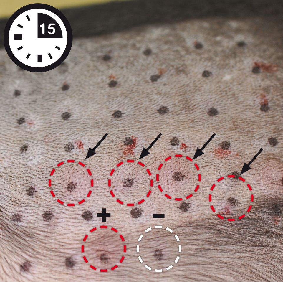
The skin site (usually the lateral thorax) is gently shaved (with the size depending on the number of allergens to be used) but should not be scrubbed or washed. Individual test sites are marked with a waterproof marker placed at least 2 cm apart, and a small volume (typically 0.05 mL) of each test concentration injected intradermally (Figure 3a). A skin bulge should appear; if absent, the allergen has been applied too deeply (subcutaneously) and the injection should be repeated.
The reactions are evaluated after 15-20 minutes, with any wheal and erythema formation at each site compared to the positive and negative controls (Figure 3b) and scored, from 0 (equal to the negative control) to 4 (equal to the positive control). Any reaction of 2 or greater is regarded as positive. Although the assessment can be done objectively (by measuring the diameter of the reaction) no definitive benefit has been noted for this option [6] and the author prefers to simply assess the reactions subjectively.
Adverse reactions to the test are rare; if they do occur it is predominantly during the actual procedure, usually as an intense pruritus at the injection site (local hypersensitivity reaction) which can be alleviated by a short course of topical glucocorticoids or systemic anti-inflammatory or anti-pruritic treatment. Rarely, other events such as anaphylaxis (generalized itching, vomiting, diarrhea or even collapse) can develop, and should be addressed appropriately.
* IDT: Intradermal testing
** PNU: Protein Nitrogen Units
*** ASIS: Allergen-specific IgE serology


Ana Rostaher
Allergen-specific IgE serology (ASIS)
In vitro ASIS is widely used in veterinary medicine as it offers several advantages over IDT. These include elimination of life-threatening risks for the patient (related to sedation or anaphylactic reactions), convenience (no hair clipping, no restraint, short duration) and a lower likelihood of prior or current drug therapy adversely influencing results [9]. Various tests are available, either as solid phase RAST or ELISA methods (the latter being the most frequently used) or as a liquid-phase immunoenzymatic assay [9]. When first introduced, these IgE tests demonstrated some disadvantages, especially poor specificity. Various improvements, particularly with the development of appropriate anti-canine IgE detection reagents, has improved their diagnostic accuracy [11]. Other limitations of ASIS are the potential for inter-and intra-laboratory variability and cross-reactivity [12]. Furthermore, recent data show that the presence of IgE antibodies against cross-reactive carbohydrate determinants (anti-CCD antibodies) may be partially responsible for false positive results, especially with pollens [13]. Blocking anti-CCD antibodies has resulted in a markedly improved correlation between IDT and ASIS in dogs [12] and a notable decrease in positive reactions to pollen allergens in cats [14]. Clinically relevant is the fact that the results obtained with ASIT do not appear to depend on the choice of ASIS methodology [9] – and as noted above, ASIT efficacy is comparable whether the choice of allergens is based on IDT or ASIS results. Because of this, ASIS may be the preferred diagnostic choice for first-line clinicians where IDT is not an option, either in-house or via referral to a specialist center.
Other testing options
Skin-prick testing is the method of choice for detection of Type I hypersensitivity in human atopic dermatitis, for several reasons; low allergen costs (glycerinated allergens tend to be stable for prolonged periods of time), rapid interpretation of results, absence of side effects, and high specificity [15]. It is also said to be significantly less painful.
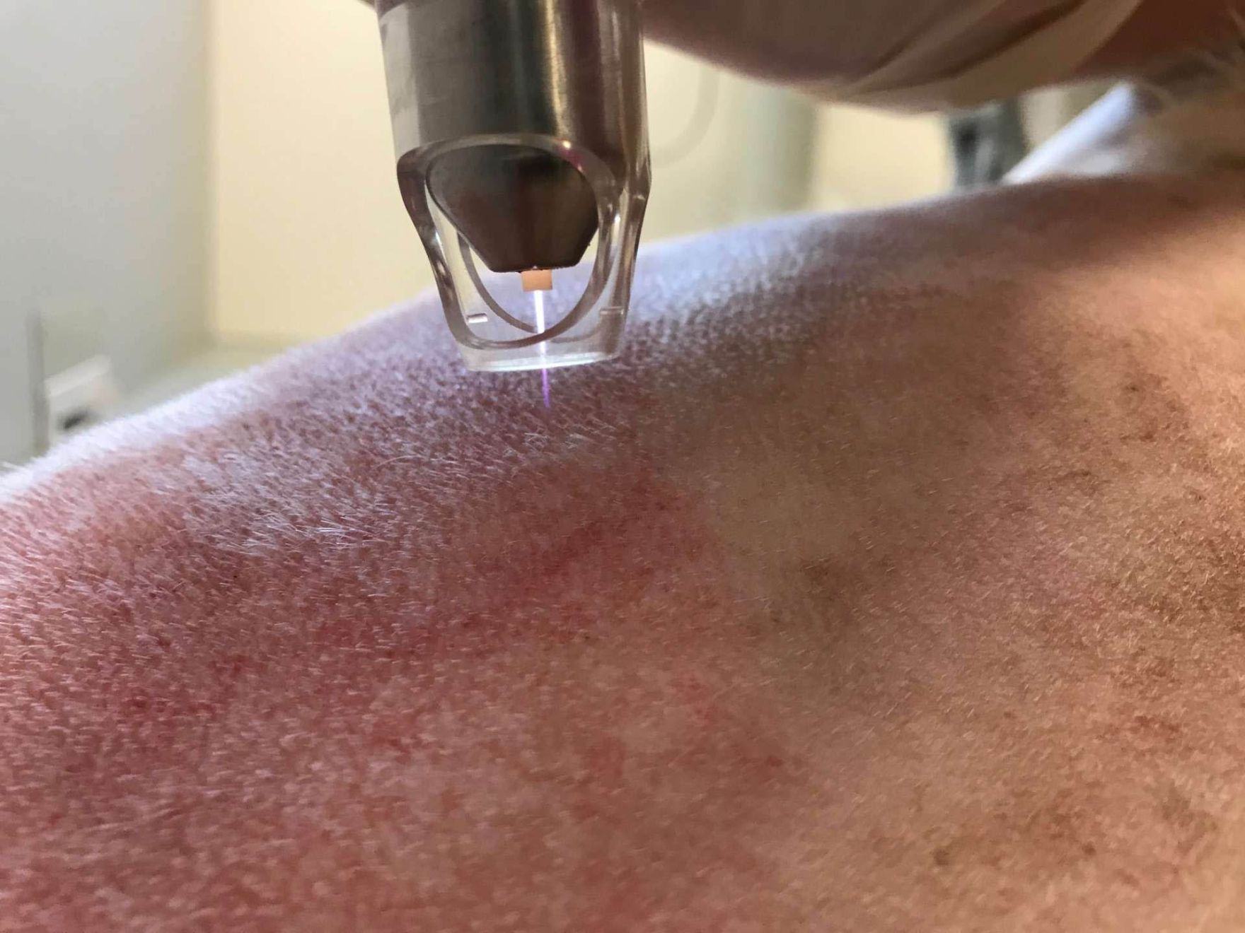
One report on prick testing in veterinary allergology dates back to the 1990s [16], but it concluded that the method was inferior to IDT in terms of result interpretation, and no further attempts were made to bring this test into clinical practice. However, within the last few years renewed clinical and scientific interest has developed to assess the benefits of this diagnostic tool in dogs and cats. In one study the test was performed in 20 healthy dogs with 8 different environmental allergens [17], with no signs of pain or discomfort noted during the simple procedure (which took on average 5 minutes, including hair clipping and allergen application). The intensity of positive results ranged from 3-12 mm (median 9 mm), but this study only assessed threshold values in healthy dogs. A similar study assessed the sensitivity and specificity of this method on 11 common environmental allergens in both non-allergic dogs and dogs with spontaneous atopic dermatitis [18]. The sensitivity was estimated to be 66% (the offending allergens could be identified in 3/5 dogs, with false negative results in the other two dogs) and 100% specificity (no dog had false positive results). Although yet to be validated in veterinary allergology, such studies suggest that prick testing might in future be a practical, accurate method that could be used as an important adjunct diagnostic for CAD. The author currently uses this test mainly to verify severe hypersensitivity reactions to Hymenoptera (e.g., bees and wasps) venom [19], with the procedure shown in Figure 4.
Lastly, various saliva- and hair-based assays for the diagnosis of adverse food reaction (AFR) and/ or environmental allergies are now available in some countries. However, recent studies in dogs showed a lack of sensitivity and specificity for any of these tests [20] [21] [22], and so their use is discouraged at least for now.


Conclusion
The diagnosis of atopic dermatitis can only be made on the basis of data derived from the patient’s history, clinical examination and by ruling out other differential diagnoses. No laboratory test can diagnose canine atopic dermatitis and therefore its over-utilization should be discouraged to limit misdiagnosis. Identification of the causative allergen in atopic dermatitis is the essential last step in the work-up, significantly influencing the long-term management and quality of life of the patient.References
- Hillier A, Griffin CE. The ACVD task force on canine atopic dermatitis (I): incidence and prevalence. Vet Immunol Immunopathol 2001;81:147-151.
- Nuttall TJ, Marsella R, Rosenbaum MR, et al. Update on pathogenesis, diagnosis, and treatment of atopic dermatitis in dogs. J Am Vet Med Assoc 2019;254:1291-1300.
- Hill PB, Moriello KA, DeBoer DJ. Concentrations of total serum IgE, IgA, and IgG in atopic and parasitized dogs. Vet Immunol Immunopathol 1995;44:105-113.
- Rostaher A, Dolf G, Fischer NM, et al. Atopic dermatitis in a cohort of West Highland White Terriers in Switzerland. Part II: estimates of early life factors and heritability. Vet Dermatol 2020;31:276-e266.
- Favrot C, Steffan J, Seewald W, et al. A prospective study on the clinical features of chronic canine atopic dermatitis and its diagnosis. Vet Dermatol 2010;21:23-31.
- Hensel P, Santoro D, Favrot C, et al. Canine atopic dermatitis: detailed guidelines for diagnosis and allergen identification. BMC Vet Res 2015;11:196.
- Park S, Ohya F, Yamashita K, et al. Comparison of response to immunotherapy by intradermal skin test and antigen-specific IgE in canine atopy. J Vet Med Sci 2000;62:983-988.
- Ansotegui IJ, Melioli G, Canonica GW, et al. IgE allergy diagnostics and other relevant tests in allergy, a World Allergy Organization position paper. World Allergy Organ J 2020;13:100080.
- Marsella R. Hypersensitivity disorders. In: Miller HW, Griffin CE, Campbell KL, eds. Muller & Kirk’s Small Animal Dermatology. 7th ed. St. Louis Missouri: Elsevier Mosby, 2013;363-431.
- Hillier A, DeBoer DJ. The ACVD task force on canine atopic dermatitis (XVII): intradermal testing. Vet Immunol Immunopathol 2001;81:289-304.
- Stedman K, Lee K, Hunter S, et al. Measurement of canine IgE using the alpha chain of the human high affinity IgE receptor. Vet Immunol Immunopathol 2001;78:349-355.
- Gedon NKY, Boehm T, Klinger CJ, et al. Agreement of serum allergen test results with unblocked and blocked IgE against cross-reactive carbohydrate determinants (CCD) and intradermal test results in atopic dogs. Vet Dermatol 2019;30:195-e161.
- Piccione ML, DeBoer DJ. Serum IgE against cross-reactive carbohydrate determinants (CCD) in healthy and atopic dogs. Vet Dermatol 2019;30:507-e153.
- Mohammaddavoodi A, Panakova L, Christian M, et al. Prevalance of immunoglobulin E against cross-reactive carbohydrate determinants (CCD) and impact of a blocker in seasonal allergy tests. Tierarztl Prax Ausg K Kleintiere Heimtiere 2020;48:404-409.
- Bousquet J, Heinzerling L, Bachert C, et al. Practical guide to skin prick tests in allergy to aeroallergens. Allergy 2012;67:18-24.
- Ballauf B. Vergleich von Intrakutan- und Pricktest in der Allergiediagnostik beim Hund. Tierärztl Prax 1991;19:428-430.
- Carnett MJH, Plant JD. Percutaneous prick test irritant threshold concentrations for eight allergens in healthy nonsedated dogs in the USA. Vet Dermatol 2018;29:117-e147.
- Carmona-Gil AM, Sanchez J, Maldonado-Estrada J. Evaluation of skin prick-test Reactions for allergic sensitization in dogs with clinical symptoms compatible with atopic dermatitis; a pilot study. Front Vet Sci 2019;6;448.
- Rostaher A, Mueller R, Meile L, et al. Venom immunotherapy for hymenoptera allergy in a Dog. Vet Dermatol 2021;32(2):206-e52.
- Coyner K, Schick A. Hair and saliva test fails to identify allergies in dogs. J Small Anim Pract 2019;60:121-125.
- Lam ATH, Johnson LN, Heinze CR. Assessment of the clinical accuracy of serum and saliva assays for identification of adverse food reaction in dogs without clinical signs of disease. J Am Vet Med Assoc 2019;255:812-816.
- Vovk LU, Watson A, Dodds WJ, et al. Testing for food-specific antibodies in saliva and blood of food allergic and healthy dogs. Vet J 2019;249:89-89.

Ana Rostaher
Vet.Med., Dip. ECVDSwitzerland
Dr. Rostaher graduated from the Slovene veterinary school in 2002 and spent four years working in small animal practice whilst finishing an internship at Vienna’s Veterinary College. She then undertook a dermatology residency in Munich and also completed a research externship on feline hair follicle disorders. She achieved her ECVD Diploma in 2011 and is currently employed at the Vetsuisse Veterinary Faculty as senior clinician. Dr. Rostaher has authored over 100 publications on various aspects of dermatology and was also a committee member of both ESVD and ECVD, having recently served as president of the Slovenian Dermatology Study Group.