Introduction
The term "adverse food reaction" (AFR) refers to any abnormal clinical reaction resulting from the ingestion of food or food additives, and can be categorized as either toxic or non-toxic in nature [1][2]. The first type is caused by substances that are natural food components, or that are present after food preparation or contamination; they can occur in any individual and are dose-dependent. Non-toxic adverse food reactions, in contrast, depend on the susceptibility of the individual, and are classified as either food intolerances (i.e., non-immune-mediated) or food allergies (i.e., immune-mediated) (Figure 1).
Food intolerances, which (at least in humans) account for most AFRs, include enzymatic reactions and those resulting from the pharmacological properties of food [1][3]. Food allergies are abnormal immunological responses to ingested food, and are specific and reproducible [4]. In humans these responses may be IgE mediated, non-IgE mediated or mixed. IgE-mediated responses are the most studied (and best defined in literature), and include urticaria and angioedema, rhinoconjunctivitis, laryngeal edema, dysphonia, oral allergic syndrome, gastrointestinal signs, systemic anaphylaxis and exercise-induced anaphylaxis [5]. The group of non-IgE-mediated disorders includes dermatitis herpetiformis, enterocolitic syndrome, colitis, proctitis, gastroesophageal reflux, celiac disease and pulmonary hemosiderosis. The mixed hypersensitivity category includes atopic dermatitis, esophageal and gastrointestinal eosinophilic disorders, and asthma. In dogs it is more difficult to make this differentiation, both because there are insufficient studies on the pathogenetic mechanisms of AFR and because clinical manifestations are not as heterogeneous as in humans, and the clinical picture often overlaps. In addition, there is no accurate test for their diagnosis and differentiation, so the more general term "adverse food reactions" is used to refer to this group of canine allergies.
Incidence, prevalence and predispositions
AFR is the third most common canine skin allergy (after flea bite hypersensitivity (FBH) and canine atopic dermatitis (CAD)). It is estimated that approximately 25-30% of dogs on a dietary elimination trial show a response to the altered diet and thus have an adverse reaction to food. A recent systematic review reported that the prevalence of AFR varied depending upon the type of diagnosis made: 1-2% of any diagnosis; 0-24% among skin diseases; 9-40% of dogs with pruritus; 8-62% of dogs with any skin allergic condition; and 9-50% of dogs with skin lesions suggestive of atopic dermatitis [6]. However, diagnosis of an adverse skin reaction to food is only confirmed by a relapse of symptoms after provocation with the food responsible for the reaction. Not all studies involve provocation tests, so AFR may be over-diagnosed, as many animals can respond because the new diet is of higher quality, or because of other therapeutic interventions (e.g., antiparasitic, antimicrobial or shampoo treatments) given in conjunction with the diet.
Pathology and possible triggers
The pathogenetic mechanisms of AFR are not fully understood. The gastrointestinal tract is continuously exposed to foreign antigens from food, microbiota or pathogens, and while some of these antigens are harmless, others are dangerous and must be removed. A breach in the mucosal barrier promotes local inflammation and increases the interaction between the luminal antigen and mucosal immune system.
In a healthy animal, lymphocytic activation occurs only when a potentially dangerous allergen comes into contact with the immune system. Conversely, when an external but non-hazardous allergen (such as a food allergen) is captured, various mechanisms are put in place to induce tolerance. The process that inhibits lymphocytic activation is called oral tolerance, and it is now recognized that there are multiple mechanisms involved, with one of the prime determinants being the dose of antigen fed. Low doses favor the induction of regulatory T cell (Tregs), whereas higher doses favor the induction of anergy or deletion, although these processes are not exclusive and might have overlapping functionality.
Although these mechanisms are very efficient in the majority of the population, individuals may be sensitized against food because of a deficient induction of oral tolerance or a breakdown in established oral tolerance [7]. As yet it is not fully understood why these abnormal responses occur, but it is clear that the cause is multifactorial: both host and food-related factors are involved [8].
Signalment
A recent study analyzing signalment data from 825 dogs with food allergy produced useful information. The age at onset varied from a few months to 13 years, with an average of 2.9 years [9]. 22% of dogs showed the initial clinical signs within the first 6 months of age and 38% when less than a year old. The most represented breeds were the German Shepherd (13%), the West Highland White Terrier (WHWT) (11%), and Labrador and Golden Retrievers (19%), which together comprise more than 40% of all cases. Labradors and WHWT were considered to be predisposed when compared to the prevalence of these breeds in the normal population. There is no definite trend for sex predisposition, which seems to vary widely between studies, with a median female/male ratio of 0.9.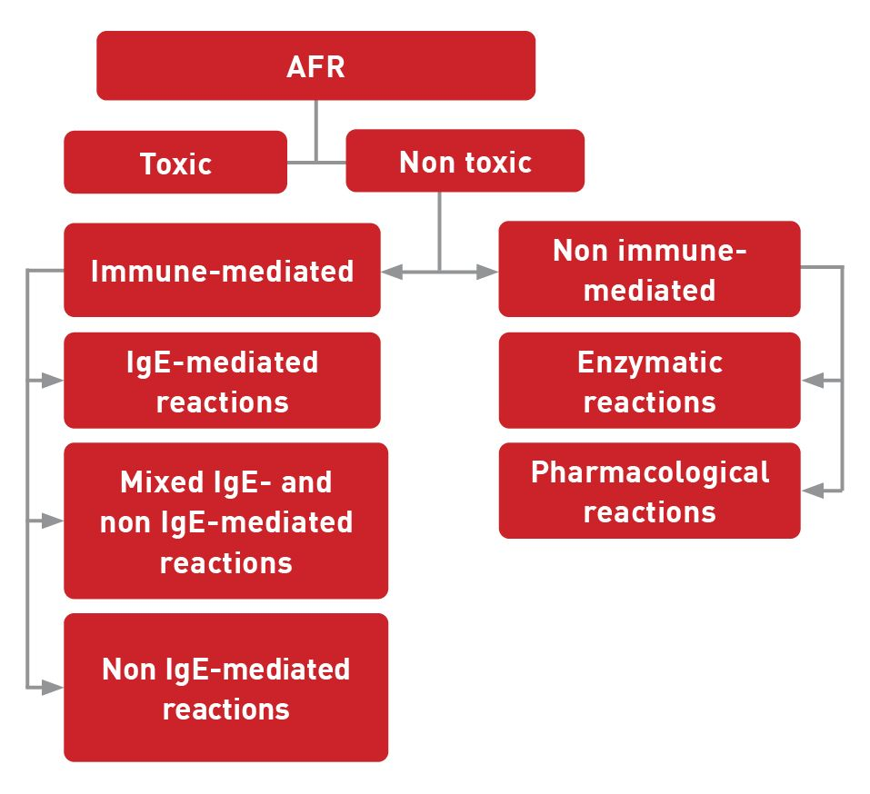
Clinical appearance
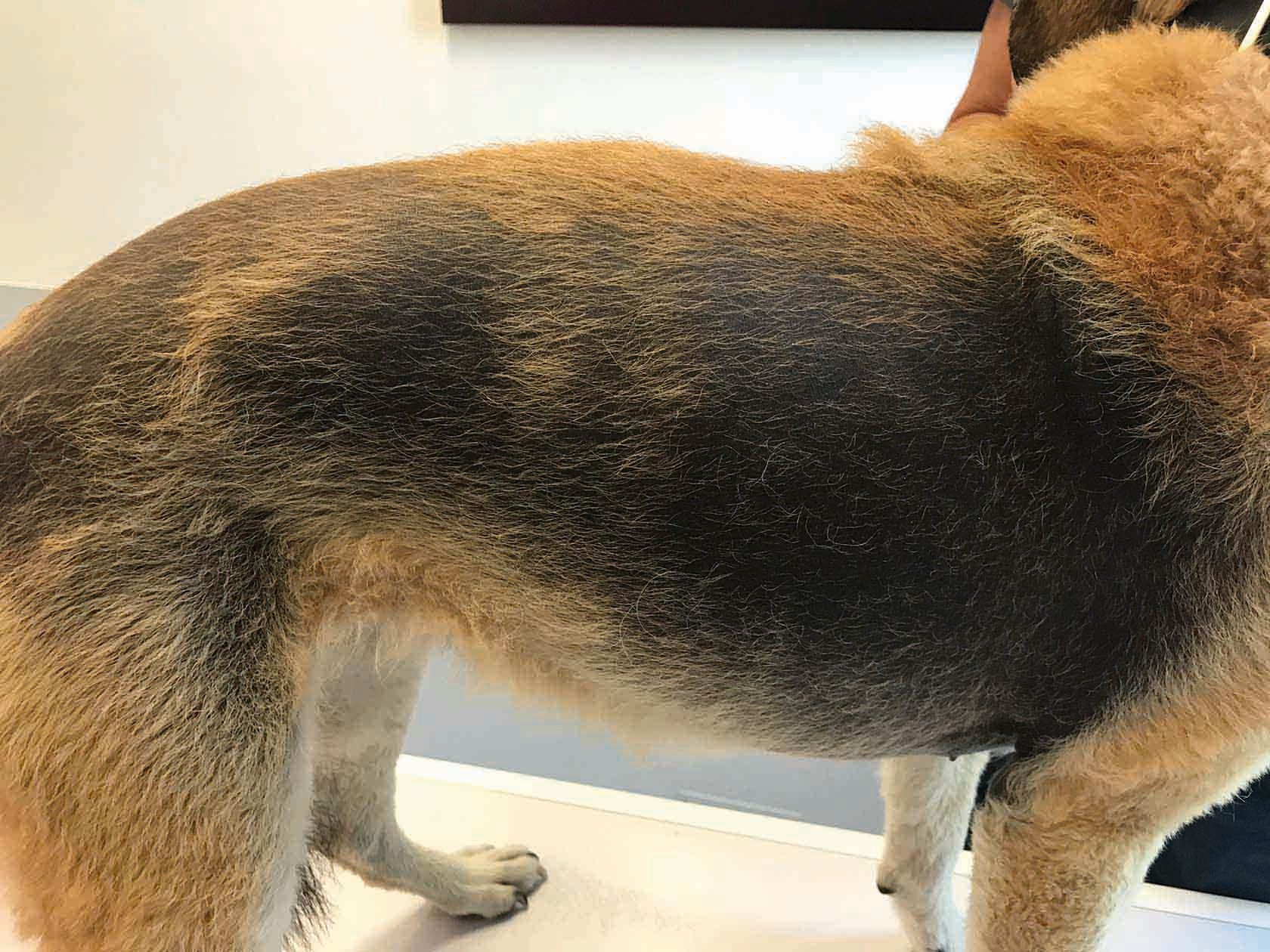
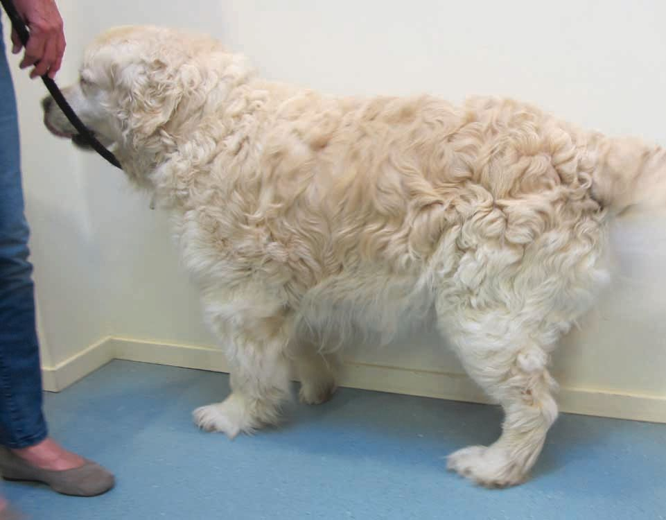
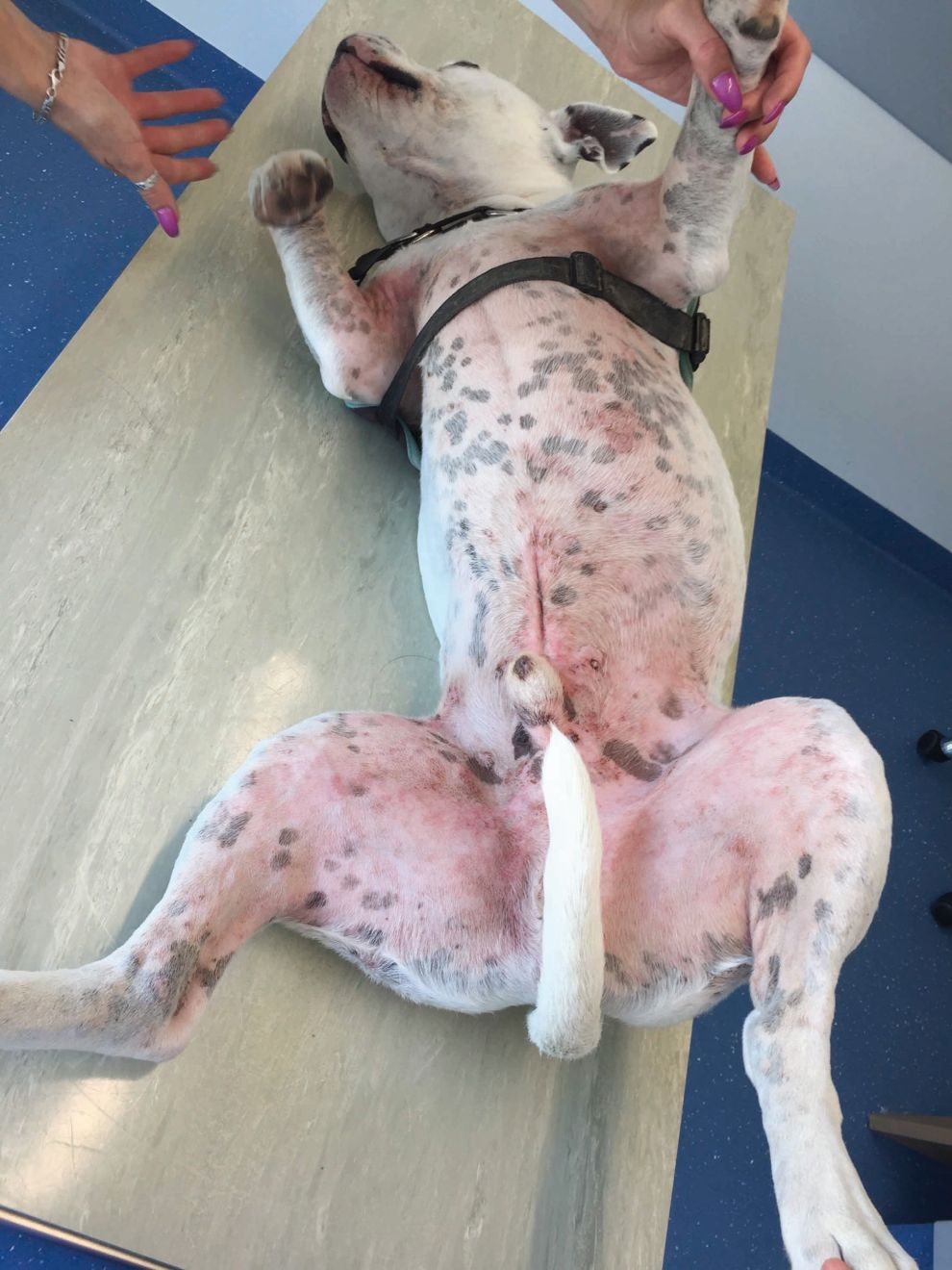
Adverse reactions to food can be difficult to diagnose due to the lack of pathognomonic signs. Non-seasonal pruritus is the most common clinical sign and often the first to appear. Itching is mainly localized in the ventral area, in particular the axillae, groin, and paws (on the palmar and/or plantar surfaces and dorsal interdigital areas). Itching of the ears is also frequently noted. A recent critical review that evaluated the dermatological signs of canine AFR suggested that approximately 50% of affected dogs demonstrate generalized pruritus (Figure 2) and that anal irritation, although reported in some individuals, is uncommon (4-25%) [10].
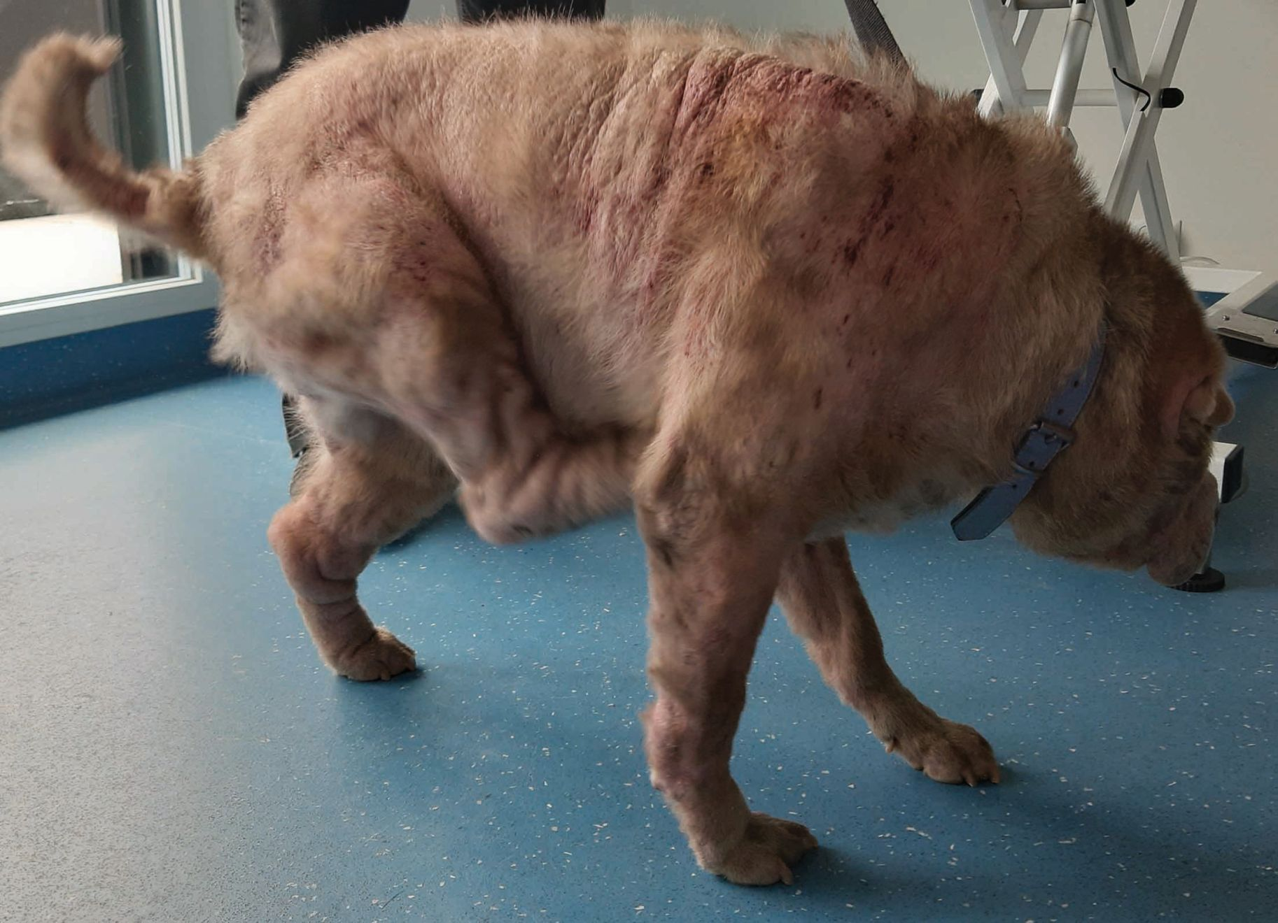
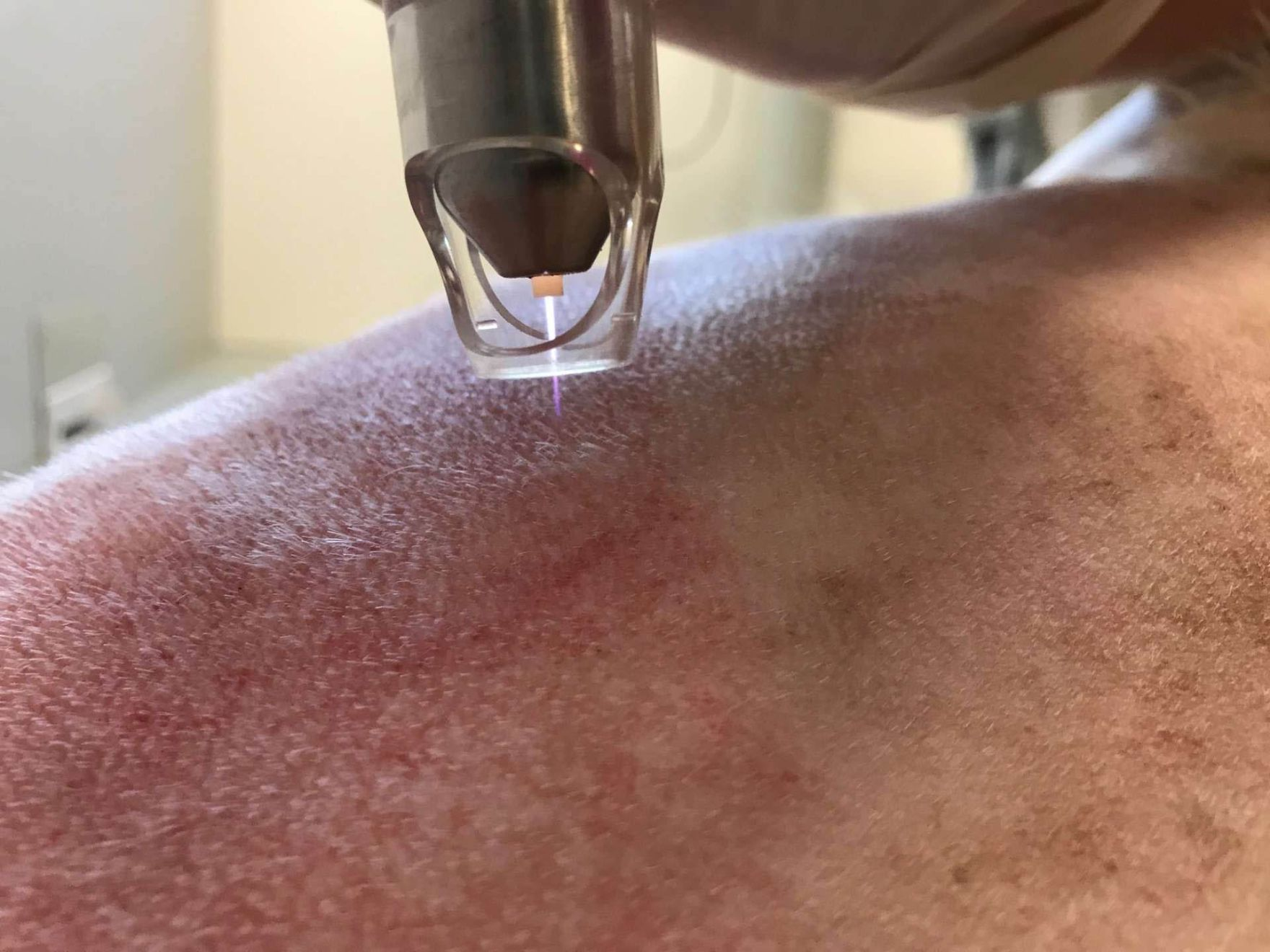
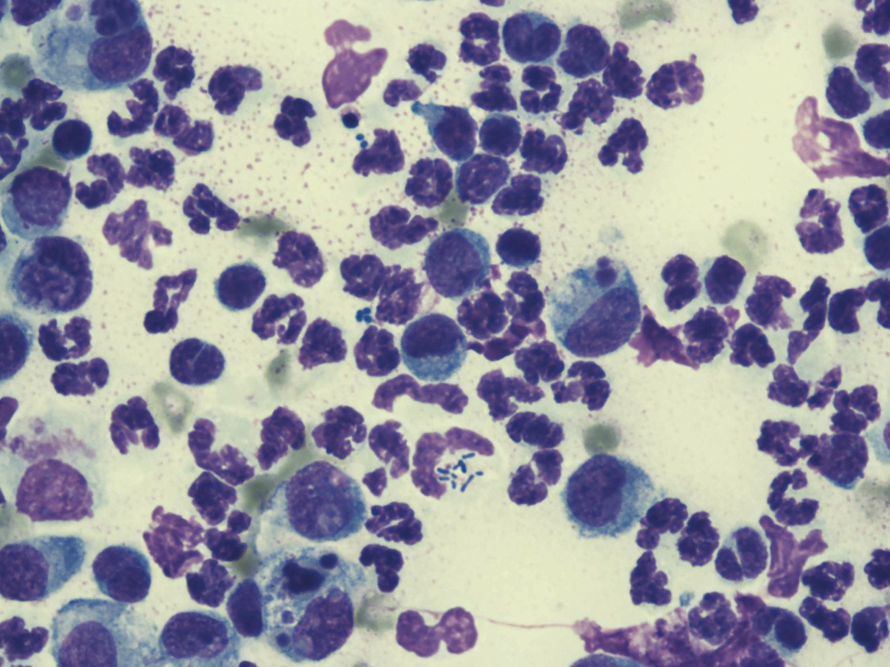
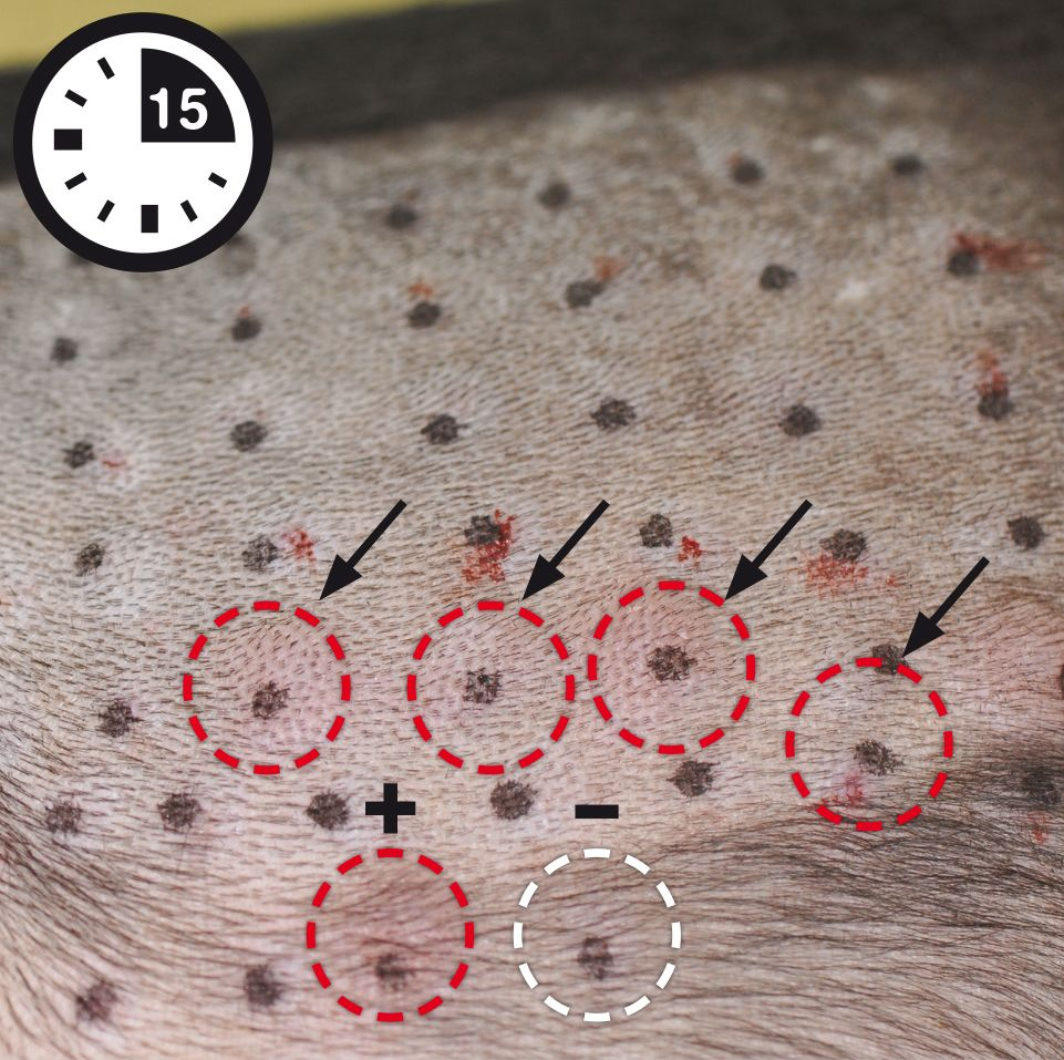
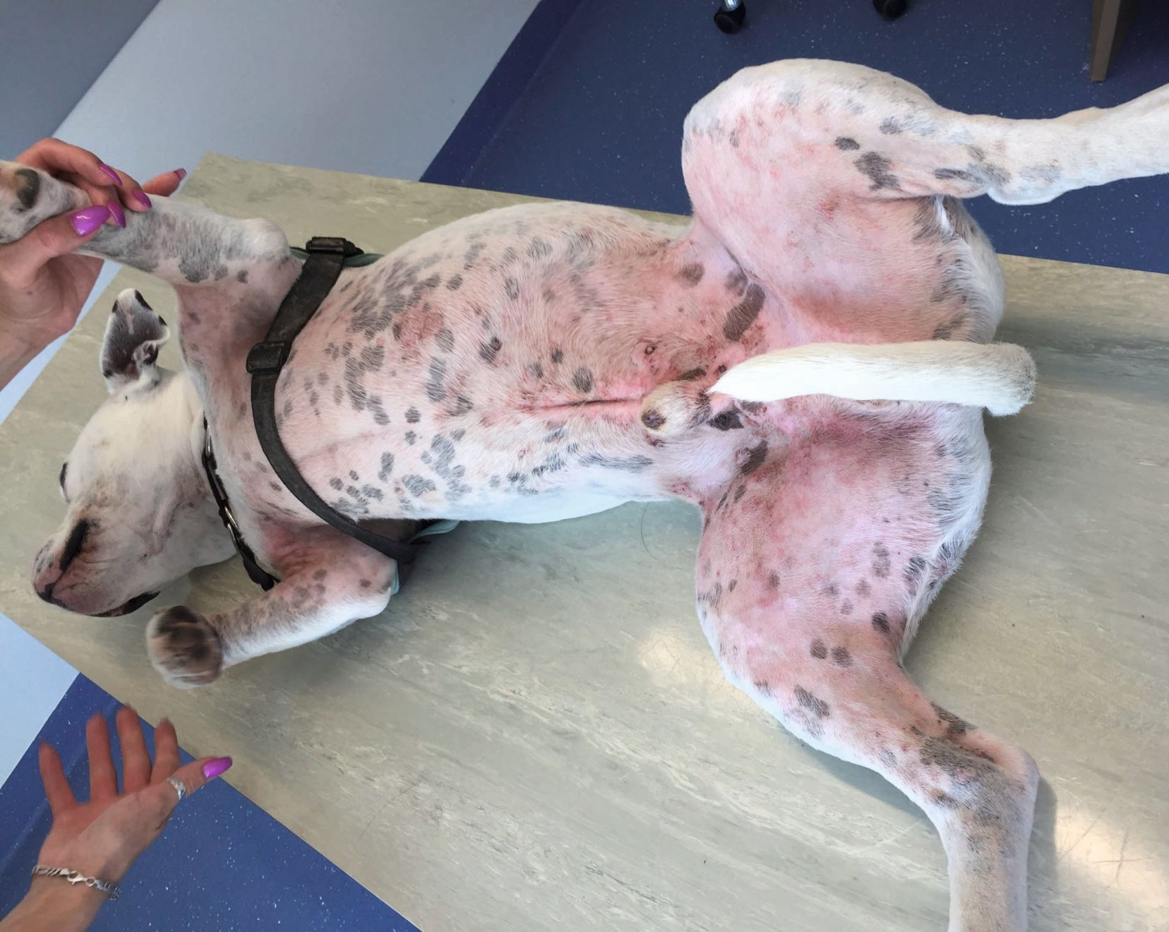
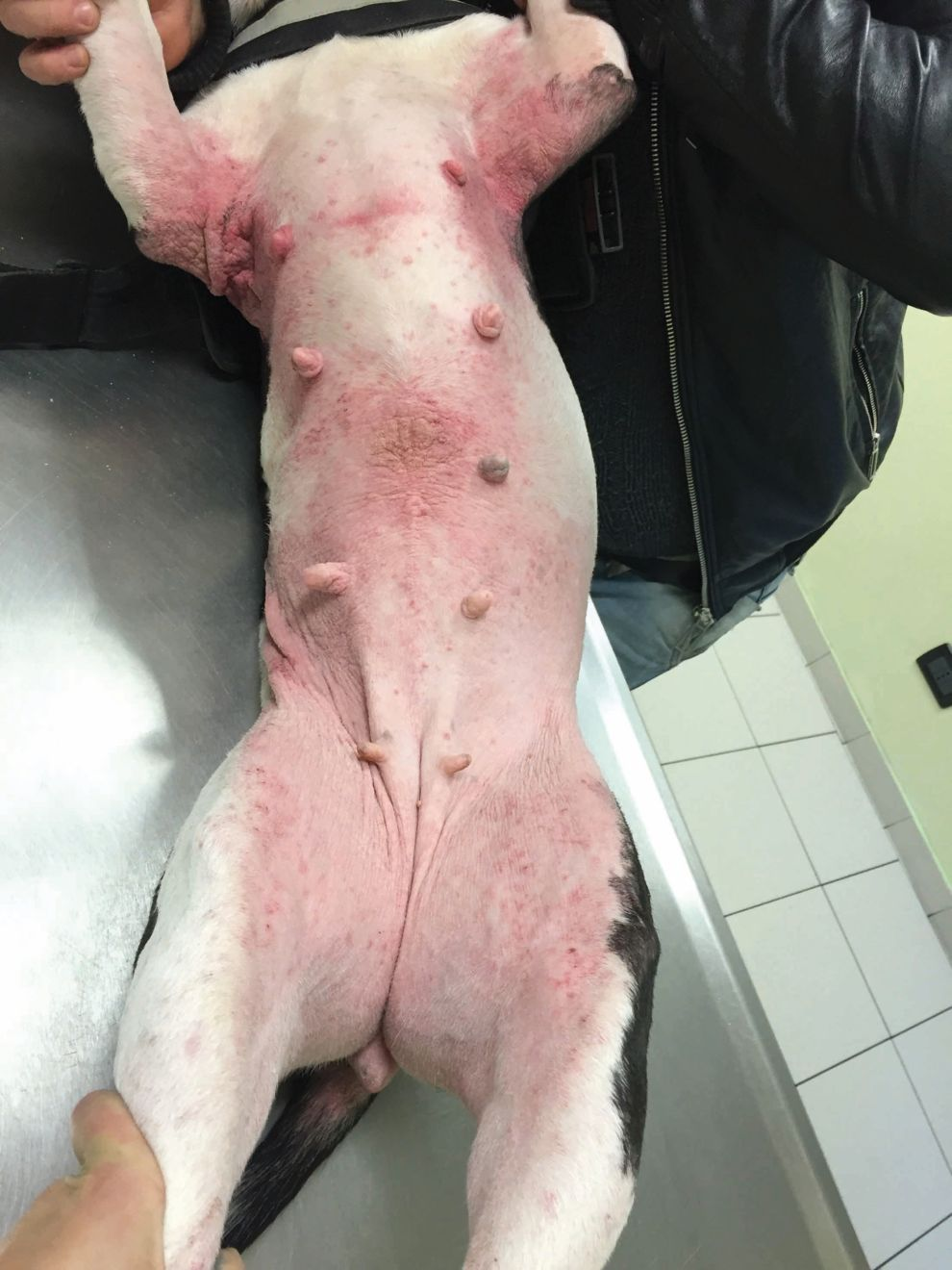
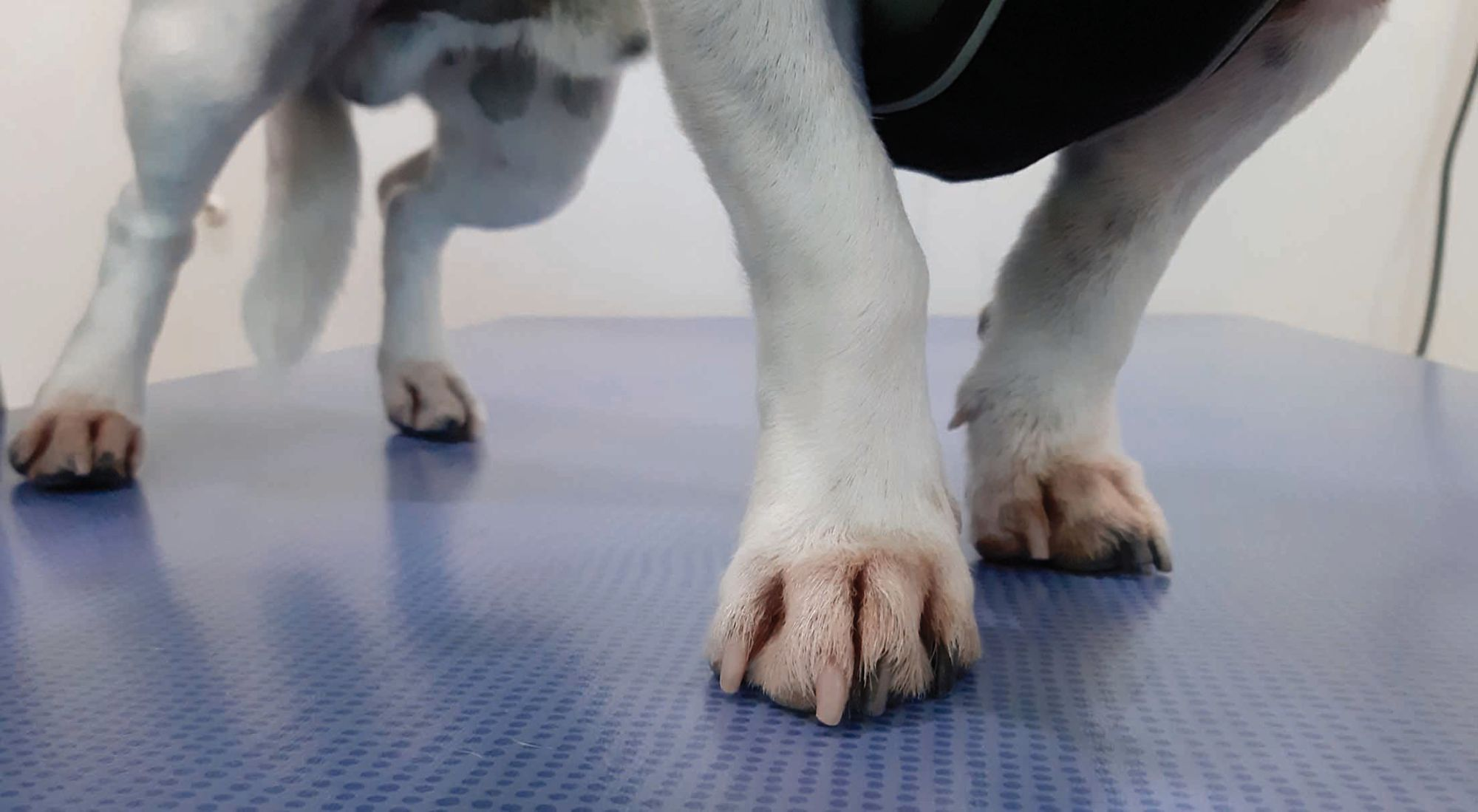
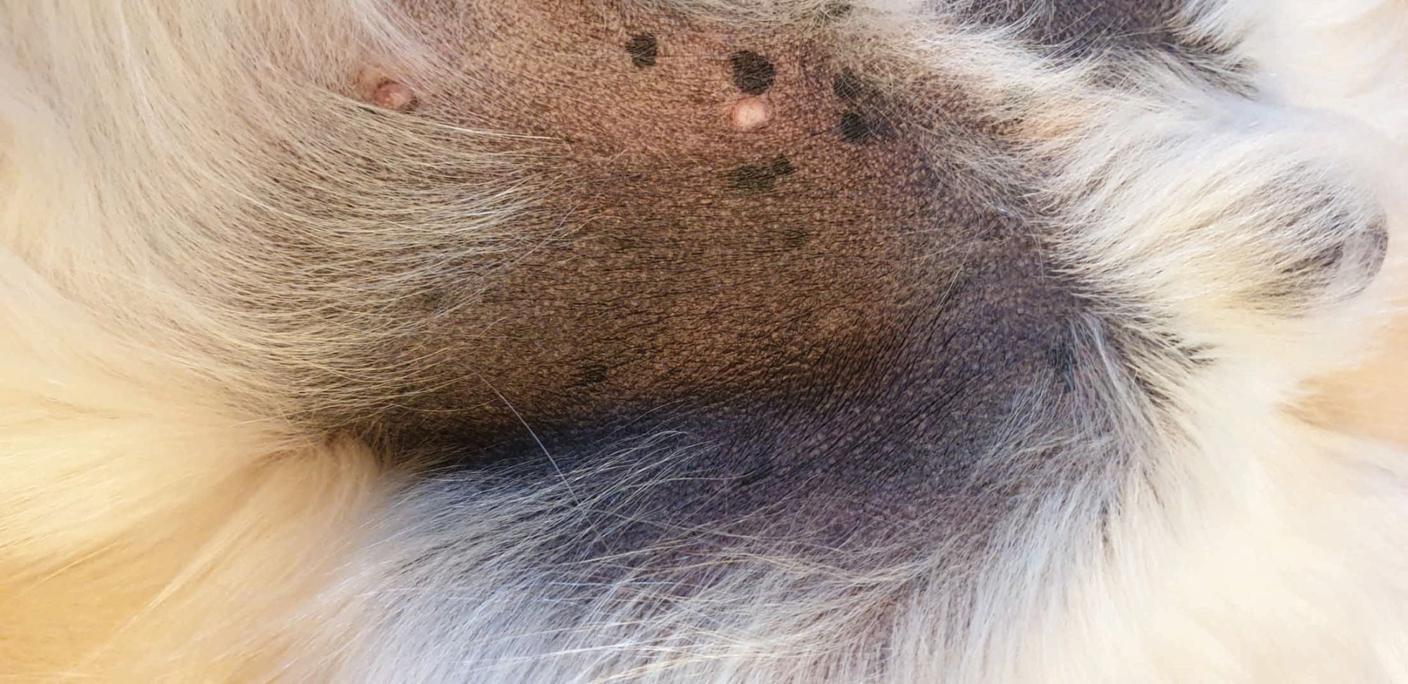
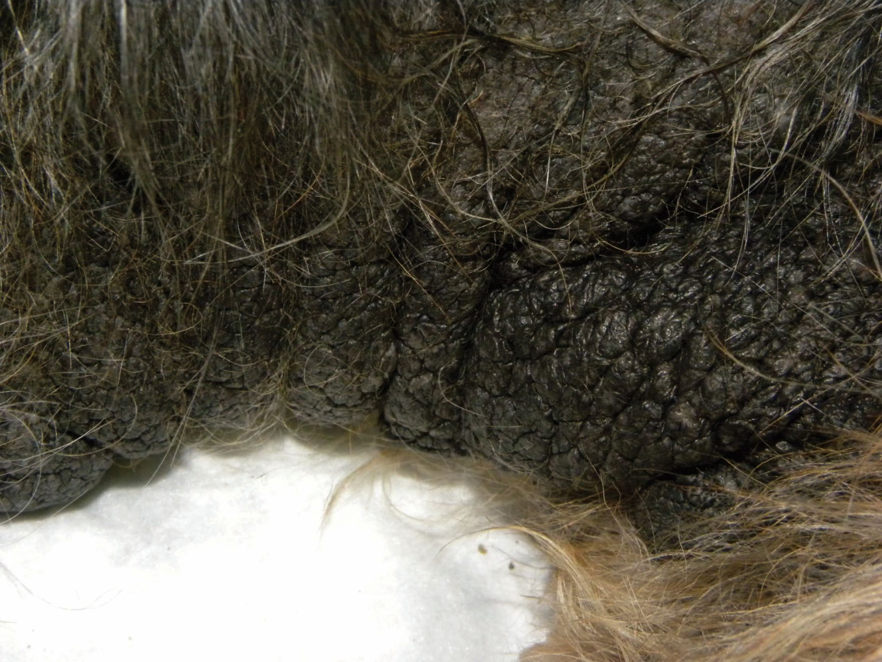
Although itching often occurs in typical areas, it is not pathognomonic, as many other skin diseases will involve the same regions, particularly other forms of hypersensitivity such as non-food-induced atopic dermatitis and FBH. Erythema and papules, with a distribution similar to that of the pruritus, are often reported as an adverse reaction to food (Figure 3), whilst other skin signs may include self-trauma caused by the dog scratching or licking itself, brownish discoloration of the hair on the paws (Figure 4), hypotrichosis, alopecia, excoriations and crusts. Over time, skin trauma causes hyperpigmentation and lichenification, and can lead to secondary skin infections (Figures 5 and 6). If not treated promptly, bacteria and/or yeasts perpetuate the inflammation (Figure 7), aggravating the dog and setting up a vicious cycle whereby the pruritic sensation leads to increased scratching and a worsening of the self-trauma.
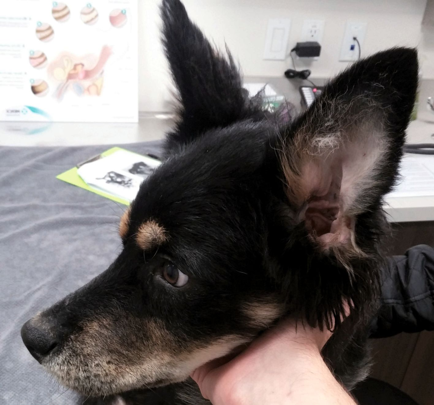
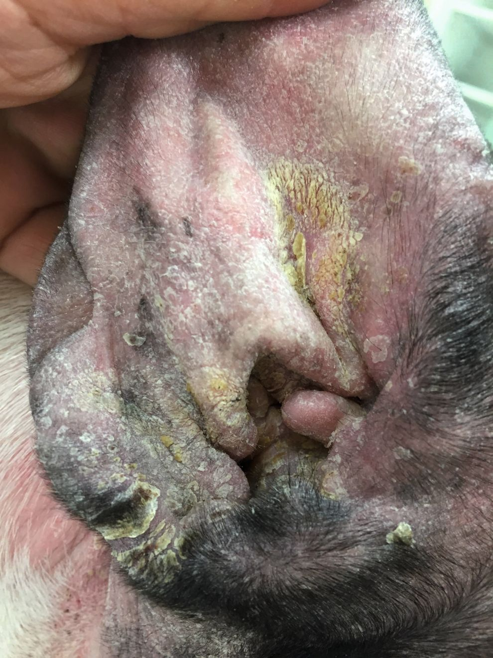
Between 13-100% of AFR cases can resemble CAD (i.e., an inflammatory, itchy skin with characteristic clinical signs), but it can also present as recurrent superficial pyoderma (in 11-70% of cases). External otitis is commonly seen (3-69%) and is often associated with pruritus (80%) but may also be the only symptom [11][12] (Figure 8). Other possible presentations include pyotraumatic dermatitis (1-9%), or – less frequently – Malassezia dermatitis, urticaria and perianal fistulae. Angioedema, urticarial vasculitis, neutrophilic leukocytoclastic vasculitis, oral allergy syndrome, erythema multiforme and interdigital furunculosis secondary to AFR have all been reported rarely.
In addition to dermatological signs, AFR may also cause gastrointestinal signs; these include chronic diarrhea and/or vomiting, soft fecal consistency or increased frequency of defecation. Abdominal pain, borborygmi and flatulence are also reported. Concurrent gastrointestinal and dermatological signs have been observed in 6-44% of affected dogs, but are not considered pathognomonic. Other, much rarer, enteropathies linked to AFR have also been reported, and are characterized by chronic intermittent or persistent diarrhea with a notable response to elimination diets.
Finally, AFR can be associated with conjunctivitis, and (rarely) respiratory disease – including bronchitis, rhinitis and chronic obstructive pulmonary disease – and even convulsions.
Diagnostic findings
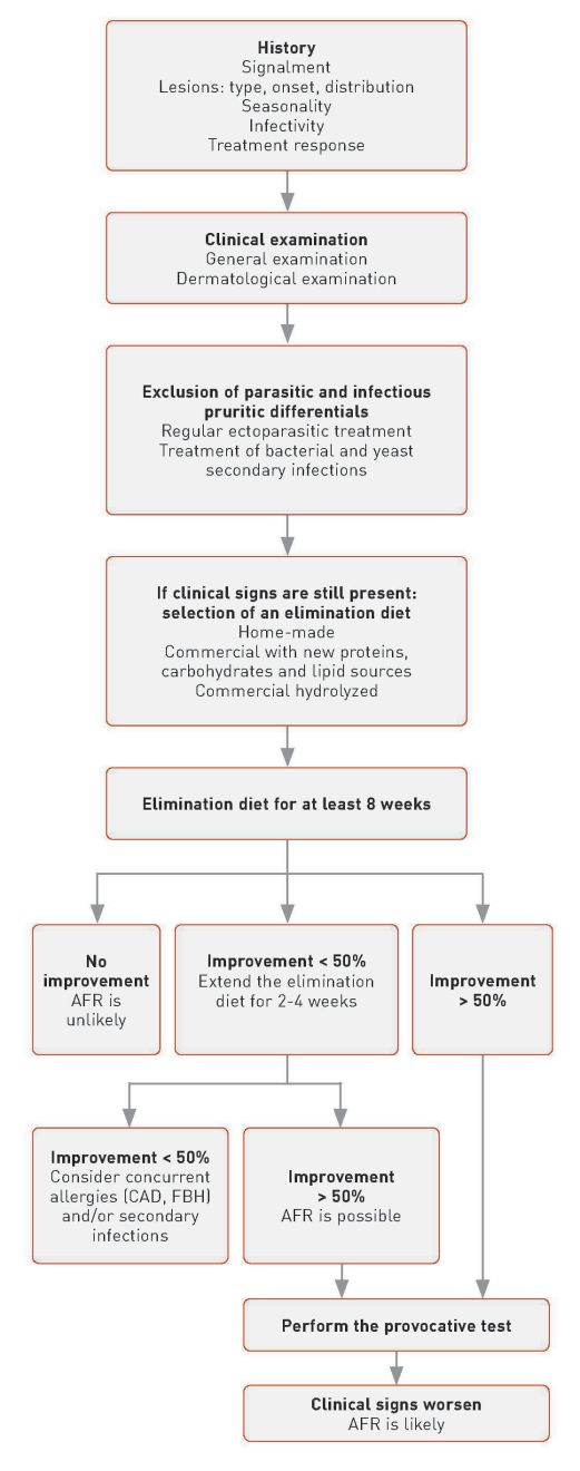
The diagnosis of AFR is based on history, clinical signs, exclusion of other pruritic diseases and a dietary trial (Figure 9). Because the signs are various and non-pathognomonic, other differentials (parasitic, infectious and allergic causes) must be considered. Ectoparasitic infestations (e.g., Sarcoptes mange) and FBH can be excluded by skin testing and ectoparasite control. Secondary bacteria and yeast infestations should first be confirmed cytologically and then appropriately treated. If signs are still present after these causes have been excluded, then an allergic etiology is likely. However, it is necessary to differentiate between AFR and CAD, since the clinical signs can be identical and there are no laboratory tests that allow a reliable differentiation.AFR is typically diagnosed following an elimination diet trial. This involves administering a foodstuff based on either a protein source novel to the dog’s immune system, or a diet based on hydrolyzed protein. Note however that commercial diets can vary in the degree of protein hydrolyzation, and the clinician should select the diet with care [13]. Some authors recommend the use of home-made recipes rather than commercial “hypoallergenic” diets because this decreases the risk of mistakenly introducing unwanted food components, but these can be problematic – for example, they can be nutritionally unbalanced, time-consuming to prepare, and expensive, especially for large breeds.
Commercial hypoallergenic diets should employ an extensively hydrolyzed protein source; although they may contain protein sources commonly eaten by the dog (e.g., chicken), an effective processing method will remove the allergenic epitopes, which prevents the immune system from recognizing the culprit allergen.
An eight-week elimination diet trial should allow diagnosis of 90% of AFR cases [14] although a recent study showed that a shorter period is possible if the pruritus and inflammation are controlled with glucocorticoids during the first 2 weeks of the trial; dogs that do not relapse after glucocorticoid discontinuation can be provocatively challenged earlier, reducing the total time period for diagnosis [15].
Dogs that respond to the restricted diet should then be challenged by either their previous diet or its individual ingredients (at least 7-14 days for each food component), to assess for any recurrence of clinical signs. Note that individual animals can be allergic to several proteins, with 40% of dogs reacting to two ingredients and 20% to three or more [16]. Only dogs that improve when given the restricted diet and then show an exacerbation of signs once re-exposed to the offending allergen(s) are definitely diagnosed as having AFR.
Control and management
There is no cure for AFR and strict avoidance of food allergens is the only way to prevent relapses. However, accidental exposures are not uncommon, and although relapses are not life-threatening, they are unpleasant and can diminish the quality of life for both dogs and their owners, and short-term intervention may be required. This can involve topical glucocorticoids, which are beneficial for localized lesions, or systemic treatment when the lesions or pruritus are generalized. The author’s preference is for either oclacitinib (0.4-0.6 mg/kg q12H PO as long as necessary to control the relapse, then discontinued) or prednisone or methylprednisolone (0.5-1.0 mg/kg PO per day either once or twice daily) [17][18][19], with the dose gradually tapered to withdrawal once remission is achieved. The latter option tends to give a more rapid improvement than cyclosporine.
When culprit allergens cannot be identified or when accidental exposures are too frequent, long-term safer therapies are to be recommended. This typically involves oral oclacitinib or cyclosporine, as glucocorticoids should be avoided in this scenario wherever possible. Oclacitinib should be given at the same dosage as for acute flares twice daily for 14 days and then once daily thereafter. Cyclosporine should be administered at 5 mg/kg q24H until clinical signs are alleviated, then tapered to a dose that maintains remission. A recently introduced alternative is lokivetmab, a caninized monoclonal antibody (mAb) which targets IL-31 [20]. Given as a single injection once a month it has been shown to produce rapid alleviation of clinical signs, with decreased pruritus within a day of administration and a lessening of lesions within 7 days [21].
Oral essential fatty acids (EFAs) are of little use when treating acute flares due to the length of time needed for any possible beneficial effect to occur, although they do offer a glucocorticoid-sparing effect if used long term. Other drugs (e.g., masitinib, recombinant canine interferon-gamma) appear to provide little or no benefit, and in any case their use is generally off-label when employed for this situation [22]. Drugs such as high-dose oral pentoxifylline, oral low-dose weekly methotrexate, and adjunctive drugs including vitamin E and antihistamines have not been studied in detail and require further proof of efficacy.
Elisa Maina
It is also important to check for bacterial and yeast infections on the skin and ears whenever acute flares are triggered. If diagnosed, topical antimicrobial shampoos and sprays or, if necessary, appropriate topical and/or systemic antibiotics should be administered following national antimicrobial treatment guidelines [18][23][24].
Finally, sublingual immunotherapy has recently been investigated as a possible treatment for canine AFR, and at least one study has shown that it can safely induce clinical desensitization [25], so in future this option may help induce tolerance, preventing dogs from accidental exposure to food-specific allergens.
Conclusion
Dogs are prevalent to adverse food reactions (AFR) and although they can demonstrate typical clinical signs in typical locations, these are unfortunately not pathognomonic, and other diseases can manifest in the same way. To complicate matters, affected dogs may also have non-food-induced atopic dermatitis and flea bite hypersensitivity, and AFR can cause other problems, either alone or along with skin lesions. Diagnosis is based on clinical history, appearance, exclusion of other differentials and an elimination diet trial. Strict food allergen avoidance is curative (although accidental exposure can cause recurrence of clinical signs, requiring symptomatic treatment), but when the culprit allergens cannot be identified, long-term medication and dietary management are necessary to prevent relapse.
Elisa Maina
DVM, PhD, Dip. ECVD
Belgium
Dr. Maina is a Diplomate of the European College of Veterinary Dermatology and also holds a PhD for research in immunology. She graduated from Milan’s Faculty of Veterinary Medicine in 2008 and went on to study dermatology with the European School of Advanced Veterinary Studies before completing a dermatology externship at the University of Florida and a residency in Italy. She currently works in a Swiss referral clinic, but also finds time to author papers for various international dermatology journals, and she serves as Chair of the ECVD Credentials Committee.
References
- Bruijnzeel-Koomen C, Ortolani C, Aas K, et al. Adverse reactions to food. European Academy of Allergology and Clinical Immunology Subcommittee. Allergy 1995;50:623-635.
- Olivry T, Mueller RS. Critically appraised topic on adverse food reactions of companion animals (7): signalment and cutaneous manifestations of dogs and cats with adverse food reactions. BMC Vet Res 2019;15:140.
- Chesney CJ. Food sensitivity in the dog: a quantitative study. J Small Anim Pract 2002;43:203-207.
- Harvey RG. Food allergy and dietary intolerance in dogs: a report of 25 cases. J Small Anim Pract 1993;34:175-179.
- Bizikova P, Olivry T. A randomized, double-blinded crossover trial testing the benefit of two hydrolysed poultry-based commercial diets for dogs with spontaneous pruritic chicken allergy. Vet Dermatol 2016;27(4):289-e70.
- Olivry T, Mueller RS, Prlaud P. Critically appraised topic on adverse food reactions of companion animals (1): duration of elimination diets. BMC Vet Res 2015;11:225.
- Favrot C, Bizikova P, Fischer N, et al. The usefulness of short-course prednisolone during the initial phase of an elimination diet trial in dogs with food-induced atopic dermatitis. Vet Dermatol 2019;30:498-e149.
- Mueller RS, Olivry T, Prlaud P. Critically appraised topic on adverse food reactions of companion animals (2): common food allergen sources in dogs and cats, BMC Vet Res 2016;12:9.
- Gadeyne C, Little P, King VL, et al. Efficacy of oclacitinib (Apoquel) compared with prednisolone for the control of pruritus and clinical signs associated with allergic dermatitis in client-owned dogs in Australia. Vet Dermatol 2014;25:512-518.
- Olivry T, DeBoer DJ, Favrot C, et al. Treatment of canine atopic dermatitis: clinical practice guidelines from the International Task Force on Canine Atopic Dermatitis. Vet Dermatol 2010;21:233-248.
- Olivry T, Foster AP, Mueller RS, et al. Interventions for atopic dermatitis in dogs: a systematic review of randomized controlled trials. Vet Dermatol 2010;21:4-22.
- Cortinovis C, Caloni F. Household food items toxic to dogs and cats. Front Vet Sci 2016;22:3-26.
- Gonzales AJ, Humphrey WR, Messamore JE, et al. Interleukin-31: its role in canine pruritus and naturally occurring canine atopic dermatitis. Vet Dermatol 2013;24:48-53.
- Michels GM, Ramsey DS, Walsh KF, et al. A blinded, randomized, placebo-controlled, dose determination trial of lokivetmab (ZTS-00103289), a caninized, anti-canine IL-31 monoclonal antibody in client owned dogs with atopic dermatitis. Vet Dermatol 2016;27:478-e129.
- Olivry T, Bizikova P. A systematic review of randomized controlled trials for prevention or treatment of atopic dermatitis in dogs: 20082011 update. Vet Dermatol 2013;24:97-e26.
- Beco L, Guaguere E, Lorente Mendez C, et al. Suggested guidelines for using systemic antimicrobials in bacterial skin infections: part 2 antimicrobial choice, treatment regimens and compliance. Vet Rec 2013;172:156-160.
- Hillier A, Lloyd DH, Weese JS, et al. Guidelines for the diagnosis and antimicrobial therapy of canine superficial bacterial folliculitis (Antimicrobial Guidelines Working Group of the International Society for Companion Animal Infectious Diseases). Vet Dermatol 2014;25:163-175.
- Maina E, Cox E. A double-blind, randomized, placebo-controlled trial of the efficacy, quality of life and safety of food allergen-specific sublingual immunotherapy in client-owned dogs with adverse food reactions: a small pilot study. Vet Dermatol 2016;27:361-e91
- Hillier A, Griffin CA. The ACVD task force on canine atopic dermatitis (X): is there a relationship between canine atopic dermatitis and cutaneous adverse food reactions? Vet Immunol Immunopathol 2001;81:227-231.
- Boyce JA, Assa'ad A, Burks W, et al. Guidelines for the diagnosis and management of food allergy in the United States. J Allergy Clin Immunol 2010;26:S1-58.
- Sicherer SH, Sampson HA. Food allergy. J Allergy Clin Immunol 2010;117:S116-S125.
- Olivry T, Mueller RS. Critically appraised topic on adverse food reactions of companion animals (3): prevalence of cutaneous food reactions in dogs and cats. BMC Vet Res 2017;13:51.
- Egawa G, Kabashima K. Barrier dysfunction in the skin allergy. Allergol Int 2018;67:3-11.
- Pabst O, Mowat AM. Oral tolerance to food protein. Mucosal Immunol 2012;5(3);232-239.
- Mueller RS, Olivry T. Critically appraised topic on adverse food reactions of companion animals (4): can we diagnose adverse food reactions in dogs and cats with in vivo or in vitro tests? BMC Vet Res 2017;13:275.