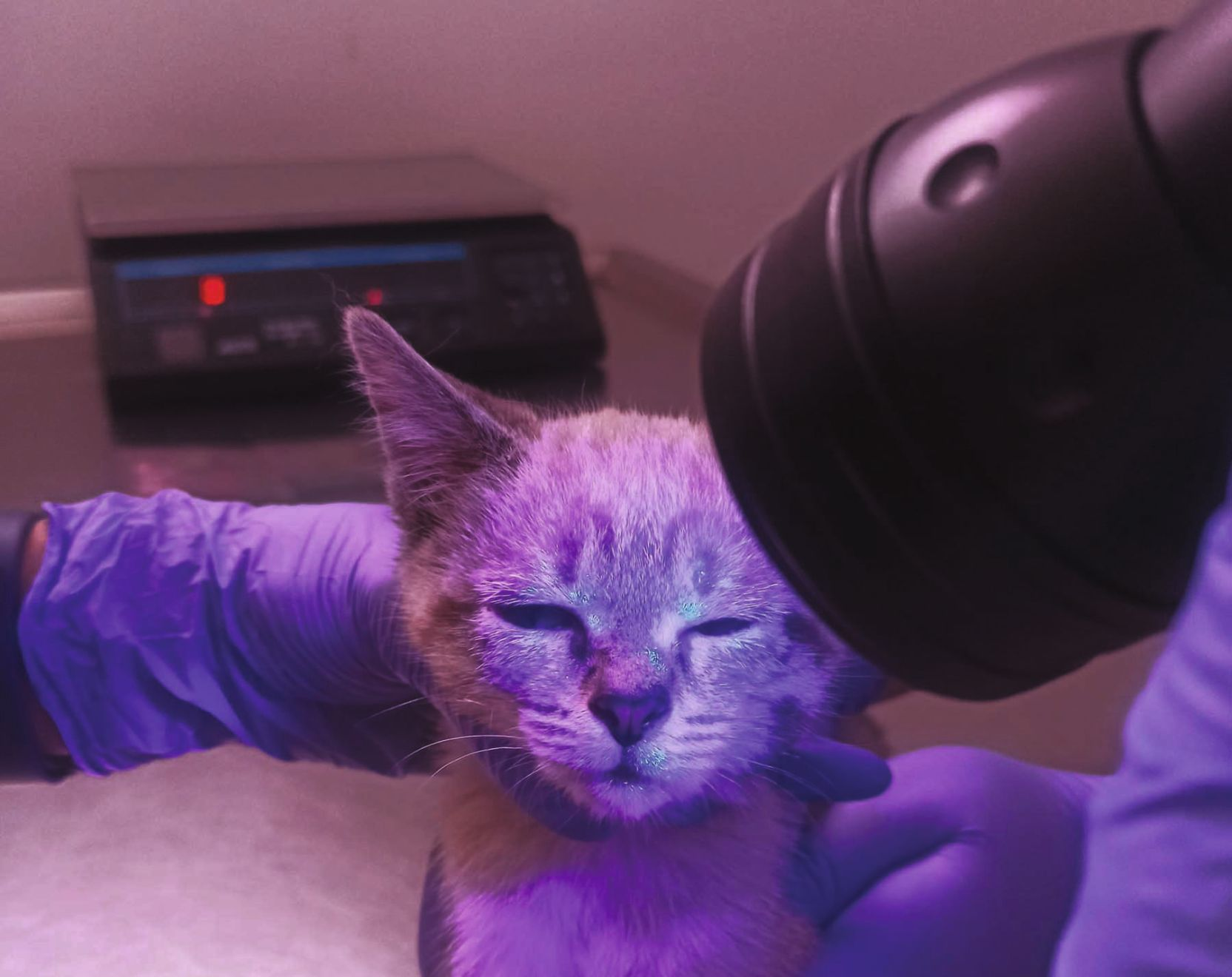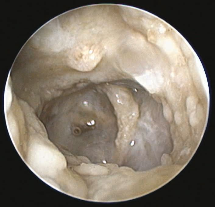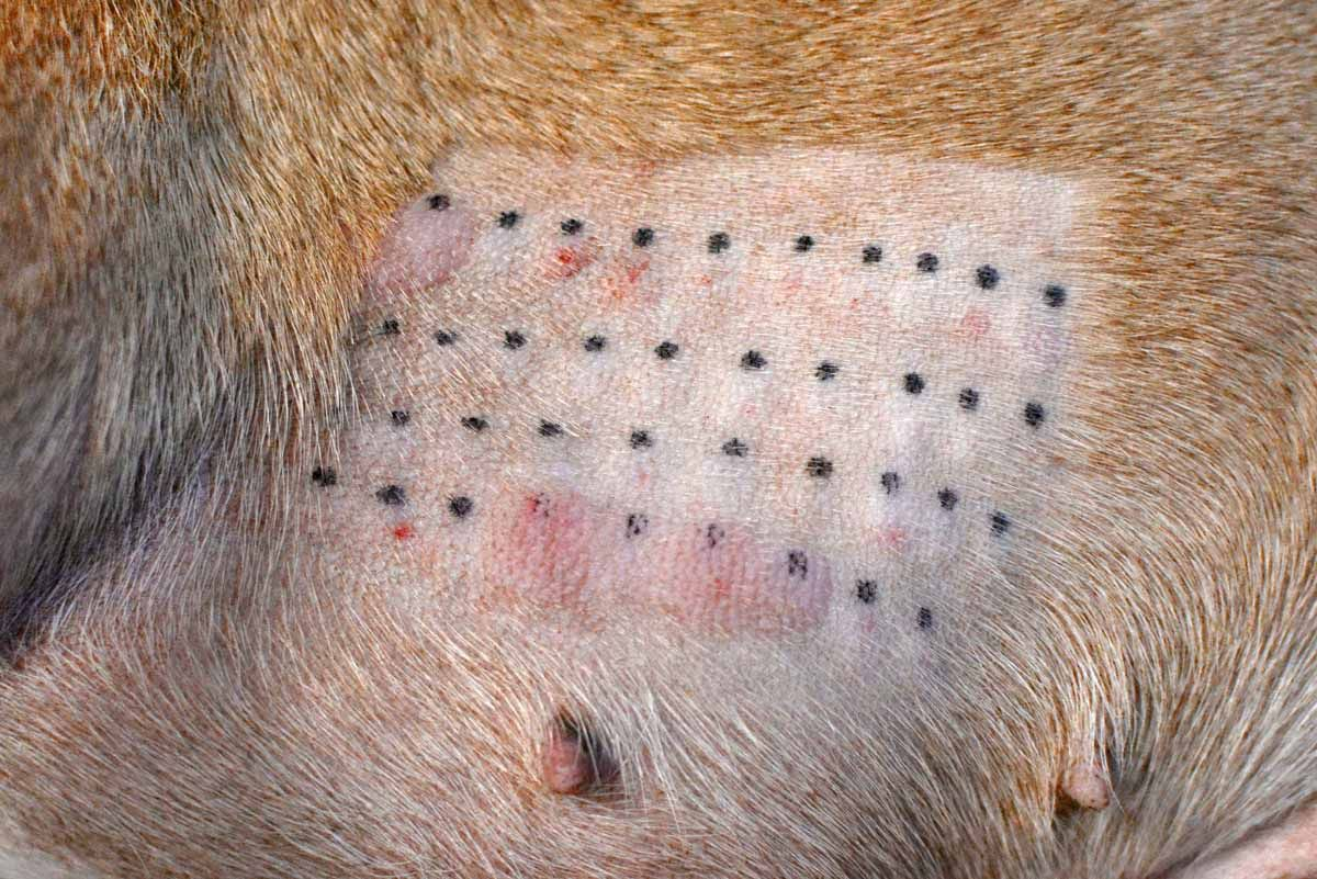
Introduction
Skin is an amazing structure and the biggest organ of the body. It serves as a vital barrier between the internal organs and the external environment, protecting an individual from foreign substances and contributing to overall health, and the skin epidermis has evolved to become a dynamic structure with homeostatic capabilities that can cope with altering external conditions. This paper will review the role of the skin barrier in relation to canine atopic dermatitis (cAD) and discuss how best to optimize its health.
Epidermal anatomy
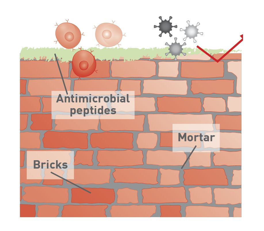
The epidermal layer of the skin consists of several tiers of corneocytes, namely (from inside to outside) the stratum basale, stratum spinosum, stratum granulosum, and stratum corneum (SC). The corneocyte is the final product of epidermal keratinization, and in dogs renewal of the epidermis takes about 22 days; new cells originating from the basal layer migrate upwards to replace the outer layer of dead cells. The cells are held together by a lipid matrix, comprising cholesterol, free fatty acids and ceramides which contribute to forming extracellular lipid-enriched lamellar membranes. A commonly used analogy to describe the skin barrier is the “brick wall theory” whereby the SC forms the “bricks”, while the lipid-enriched layers between the cells forms the “mortar” (Figure 1a) [1]. The integrity of the SC, particularly the lipid matrix, is important in maintaining the skin barrier function.
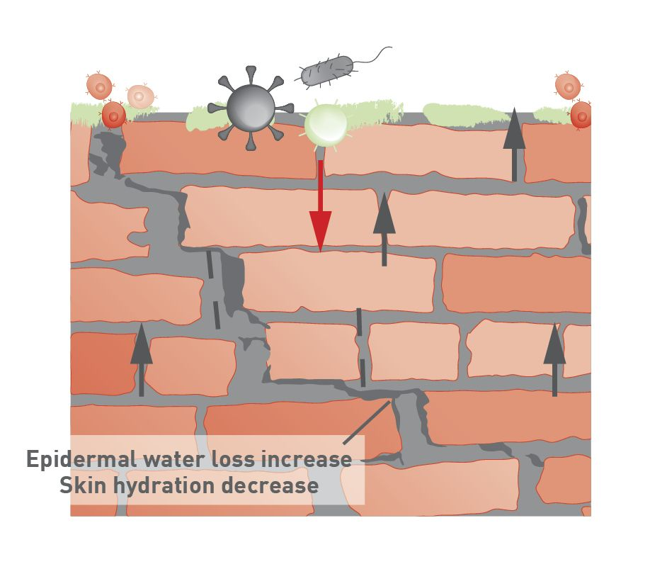
In addition to intercellular lipids, the outermost layer of the SC is coated with a variety of hydrophobic molecules, forming a protective barrier against microbes and allergens. Skin surface function and skin surface lipids have been investigated extensively in atopic dermatitis (AD) of both humans and dogs, and changes in lipid composition (free fatty acids and ceramides) in AD lesional skin and the lamellar conformation are thought to compromise the integrity of the skin barrier (Figure 1b) [2]. Although definitive evidence of the relationship between skin barrier dysfunction and the development of cAD is still lacking, a few candidate genes have been associated with defective epidermal barrier integrity in cAD, such as those encoding for the exoskeleton protein plakophilin 2 (PKP2) and filaggrin (FLG) [3]. In human medicine, transepidermal water loss (TEWL) and skin hydration (SH) are used to evaluate skin barrier function, treatment efficacy in atopy patients, and assessment of cosmetic products [4]. TEWL and SH measurements are often included in clinical trials due to their noninvasive and convenient nature, but these techniques have not yet been standardized for use in dogs.
Skin microbiota
Skin is also colonized by a diverse range of microorganisms (bacteria, Malassezia and fungi) known as the microbiota. In recent years, advances in next-generation sequencing have enabled identification of a wide range of cutaneous residents, and the microbiota can vary significantly between different body sites in the same dog. It is notable that it is susceptible to influences from factors such as topical therapy, systemic medications (especially antimicrobials), and even environmental conditions.
The exposure to a wide-spectrum microbiota during early life contributes to its adaptation to non-harmful microorganisms [5], and reflects the so-called “hygiene hypothesis” of human atopy. This was proposed in 1989 to explain the increasing prevalence of atopic conditions seen in humans; it suggests that a higher incidence of childhood infections might provide protection against the development of atopic diseases in later life. Babies tend to be born with an immune response that is Th (T-helper cell) 2 biased; this can be switched off rapidly postnatally under the influence of microbiological exposure, or can be enhanced by early exposure to allergens. Th2 cells are known to be involved with allergic responses, whereas the Th1 response is essentially mounted against infectious pathogens. Ideally, the two should be in equilibrium; a balanced Th1/Th2 pattern has been observed in infants that are less likely to develop atopic disease in later life. Unfortunately, there is no current research that supports the hygiene hypothesis in AD dogs, but research indicates a lower diversity in the skin microbiome in atopic dogs when compared to healthy dogs, which exhibit a rich diversity in their microbiota [6]. Furthermore, when dogs with AD experience acute flares, there is a temporary disruption in the balance of their microbiota (dysbiosis) due to a substantial increase in Staphylococcus spp. However, after antimicrobial therapy and during remission of lesions, TEWL is reduced, and microbial diversity is restored [7].
Canine atopic dermatitis
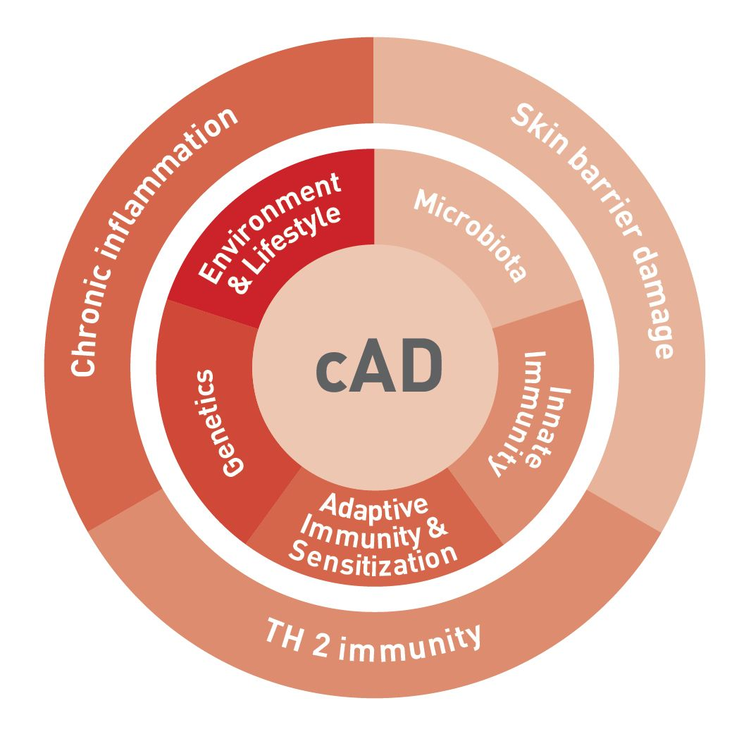
cAD is a pruritic and predominantly T-cell driven inflammatory skin disease. Its development has a multifactorial pathogenesis, involving a complex interplay between immune dysregulation, skin barrier abnormalities, genetic predisposition, environmental factors and dysbiosis (Figure 2). It is suggested that the environmental factors play a significant role in the development of cAD [3], as a dog’s lifestyle, particularly during puppyhood, has a notable influence on maturation of the immune system. As such, growing up in a rural environment, having a large number of family members, and being in contact with other animals may lower the risk of developing cAD later in life. In addition, there is also an indication that Toxocara canis infection might have a protective effect against Dermatophagoides farina (house dust mite)-induced cAD [3].
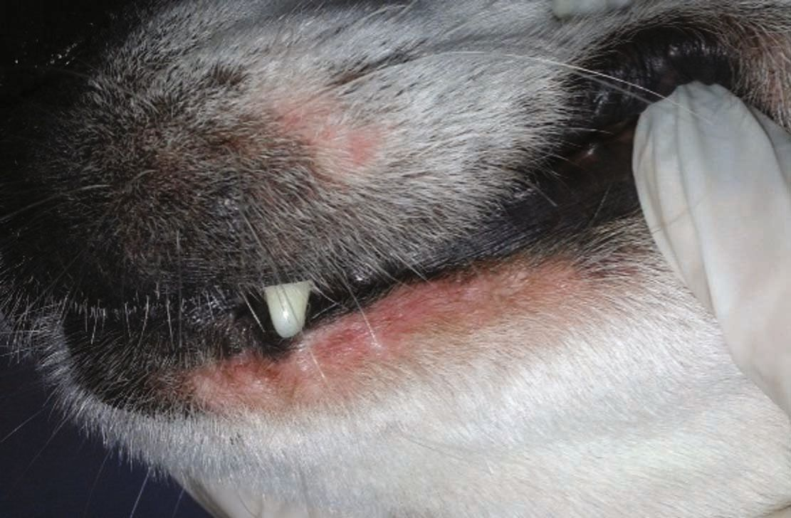
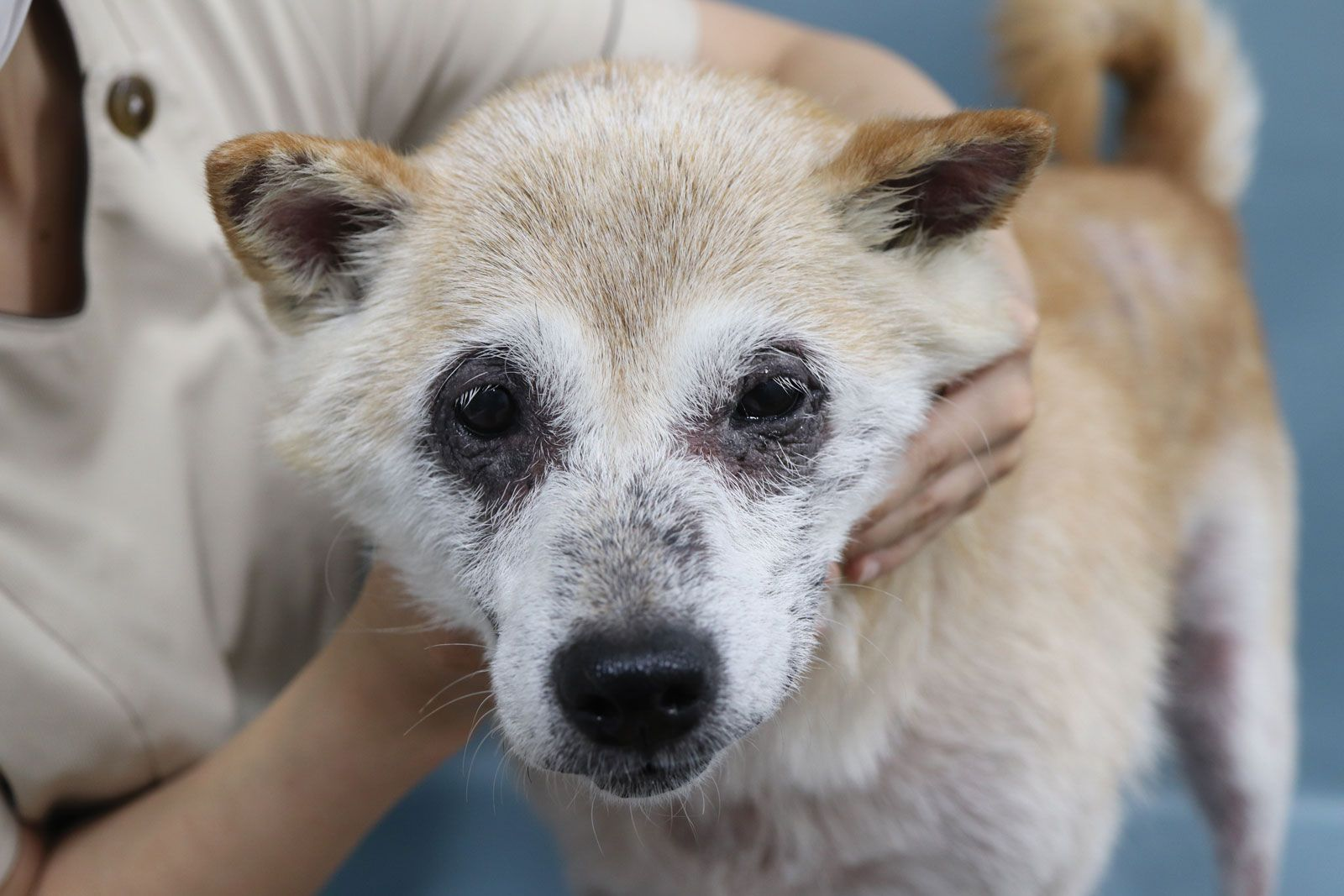
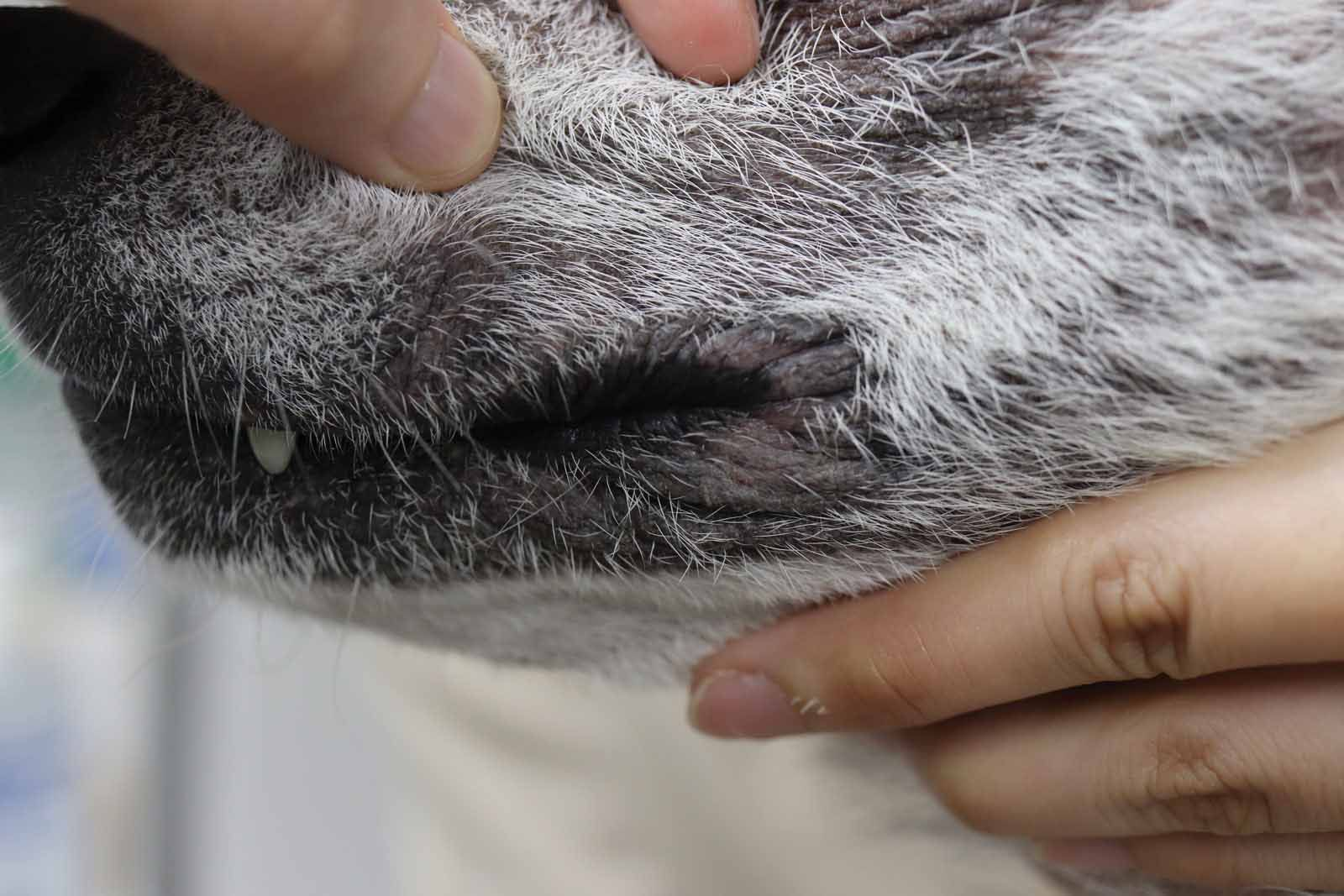
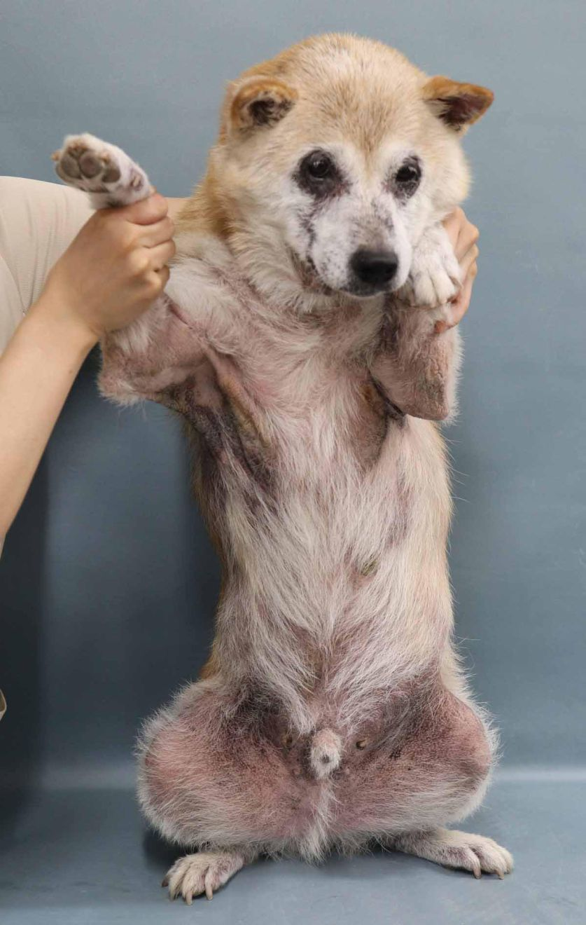
The primary clinical feature of cAD is pruritus, which can present as either a seasonal or non-seasonal pattern. Lesions are commonly found in areas such as the periocular region, around the muzzle, axillae, inguinal region, perianal area, and the extremities (Figure 3). Dogs with cAD may experience recurrent problems such as otitis externa and pododermatitis, which are commonly associated with secondary bacterial infections or an overgrowth of Malassezia. Diagnosing cAD relies on the exclusion of other pruritic skin conditions, especially those that mimic cAD, such as flea infestation/flea bite hypersensitivity, scabies, and adverse food reaction (AFR). Once ectoparasite prevention, diagnostic examinations and food trials have been successfully performed, Favrot’s criteria can be used to establish a clinical diagnosis of cAD (Table 1) [8]. For identification of cAD-related allergens, skin testing and IgE serology tests are available, although it should be realized that these tests are only needed if allergen-specific immunotherapy is considered [9].
Table 1. Favrot’s criteria for canine atopic dermatitis. If 5 or more of the criteria are met there is at least an 80% chance that AD is the cause of the pruritus.
|
Complicating factors – pyoderma
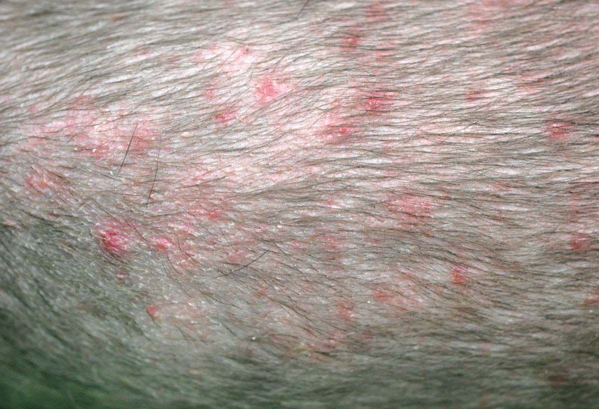
Staphylococcus pseudintermedius is one of the commensals on the canine skin and an opportunistic pathogen in pyoderma and otitis externa, commonly associated with underlying cAD and/or adverse food reactions [10]. Healthy skin has its own defense mechanisms to prevent bacterial overgrowth, such as antimicrobial peptides (AMP), beta-defensins (BDs), and cathelicidins (caths), located in the extracellular spaces of the SC [11]. The microbiota and the skin barrier work together to maintain integrity and defend against the external environment. The precise factors contributing to increased susceptibility of dogs with AD to Staphylococcus spp. infections are not fully elucidated. The diagnosis of a bacterial skin infection (superficial folliculitis and pyoderma) is based on the clinical presentation (papules, pustules or epidermal collarettes – Figure 4) and the presence of intracellular cocci on cytology. Effective management of secondary bacterial infections can be accomplished through topical treatments. Maintaining skin and coat hygiene by shampooing can be particularly beneficial for dogs with cAD, as it helps promote a healthy skin barrier. Shampooing diminishes the allergens attached to the skin surface or hair coat, preventing further irritation. Moreover, in-vitro studies suggest that shampoo or mousse preparations that include antiseptic ingredients can have residual effectiveness for up to 14 days [12], so products containing chlorhexidine, benzoyl peroxide, ethyl lactate, povidone iodine or triclosan may be beneficial in treating superficial pyoderma [13]. These should be used 2-3 times weekly, and then tapered down to once a week if the lesions resolve [13]. In addition, mupirocin and fusidic acid are recommended as topical antimicrobial agents, due to the lower risk of developing multidrug-resistant S. pseudintermedius (MRSP). Systemic antimicrobial therapy should only be considered if topical treatment proves ineffective or if the infection’s depth and location exceed the scope of topical therapy, with antibiotics selected on the basis of bacterial sensitivity tests. In general, the treatment course should be continued for 2 weeks after remission of the lesions. However, due to the global emergence of MRSP, it is highly advisable to prioritize topical treatments over repeated systemic antimicrobial therapy if the patient’s condition allows.
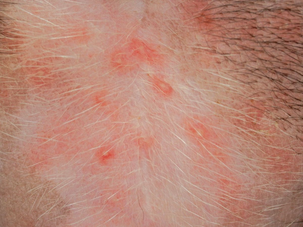

Complicating factors – Malassezia
Malassezia pachydermatis is a lipid-dependent yeast found on the skin surface. Excessive surface lipids, disruptions in the SC barrier function, and an aberrant immune reaction in cAD may contribute to overgrowth of this opportunistic pathogen [14]. Colonization in puppies occurs in a similar fashion to that of Staphylococcus spp., with transmission through licking and nursing by the mother at the very early stages of life. When the conditions are favorable, Malassezia can proliferate within the SC, producing numerous antigens and allergens [14]. These antigens can penetrate the epidermis and trigger an immune response in cAD dogs, resulting in pruritus and erythema. Malassezia tends to flourish in areas of the skin with high humidity, such as skin folds around the ears, lips, anus, axillae, inguinal region and the claws/interdigital areas. Clinical presentations may manifest as pruritus and erythema, accompanied by malodor or a greasy skin coat. There is no gold standard for diagnosing Malassezia dermatitis, but skin cytology and a compatible clinical presentation are the most effective methods; culture, histopathology and IgE tests lack substantial clinical diagnostic value.
The primary treatment options consist of antifungal agents such as topical imidazoles, clotrimazole, climbazole, and miconazole. Terbinafine and 2% chlorhexidine/2% miconazole shampoo are alternatives. However, although antiseptic ingredients can substantially diminish a Malassezia overgrowth, emollient bathing products containing cleansing oils, and emollient shampoos with a ceramide-based moisturizer have also demonstrated similar clinical efficacy [15].
Complicating factors – fleas and adverse food reactions
Flea control and elimination of food allergens are vital strategies in managing cAD, as these help to lower the threshold for pruritus, ultimately improving the patient’s quality of life. Oral adulticides are indicated for efficient year-round control of fleas, as shampooing can flush out topical products and reduce their effectiveness. Isoxazoline products have demonstrated a rapid onset of efficacy, being able to eliminate fleas within 24 hours of administration, which prevents further flea bites.
Adverse food reactions can coexist with cAD, with estimates ranging from 9-50% among dogs displaying lesions indicative of cAD. To differentiate AFR and atopic dermatitis (which is mainly due to environmental allergens) a strict 8-week food trial based on a novel or hydrolyzed protein diet must be followed. It is also advisable to consider provocative testing after the food trial in order to identify specific food allergens and subsequently remove them from the dog’s daily diet. Although the exact immune mechanism of AFR is not fully understood, a study in affected dogs that had shown significant clinical improvement on an elimination diet trial reported that 90% went on to display clinical signs again (pruritus, feet licking, face rubbing) when tested subsequently, some within a few hours of being fed the provocative diet [16].
It is suggested that environmental factors play a significant role in the development of cAD. A dog’s lifestyle, particularly during puppyhood, has a notable influence on the maturation of the immune system.
Control and management of cAD
cAD is a skin disease which cannot be cured, and it usually requires life-long management customized for the individual patient. Allergen-specific immunotherapy (ASIT) is considered the specific treatment, involving injections of increasing concentrations of environmental allergens identified by skin testing and IgE serology. The clinical effectiveness of ASIT is approximately 60% and may require 9-12 months treatment before noticeable improvement [17]. Recently, intralymphatic and sublingual immunotherapy options have been introduced as alternatives to the traditional subcutaneous injection method; these offer a more rapid induction, and the former method is needle-free [18]. However, it is essential to manage the pruritus and skin lesions continuously until clinical signs are alleviated through ASIT. Management of cAD depends on the stage of the disease – so for example, rapid intervention with acute flares, control of the chronic disease, or prevention of relapses. As pruritus is the key sign of AD and skin lesions are common secondary to this, treatment should be focused on diminishing the itch. Depending on the intensity of the pruritus, and the distribution and extent of the lesions, topical and/or systemic medication can be chosen. To control acute flares glucocorticoids (topical and systemic) and oclacitinib are considered to be the most effective because of their rapid action, although glucocorticoids are noted for potential adverse effects, which may include polyuria, polydipsia, polyphagia, increased susceptibility to infections, and iatrogenic hyperadrenocorticism. Long-term or high-dose use of glucocorticoids should be carefully monitored, with a switch to other regimes if possible. When the skin lesions have reduced to a mild level, oclacitinib is preferred for its effectiveness in managing residual pruritus and mild flares, as it diminishes the pruritogenic signaling pathway and pro-inflammatory cytokines. However, after the initial two week twice daily initiation phase, patients will often show a rebound phenomenon when tapered to once daily treatment [19]. In order to prevent this, topical hydrocortisone aceponate can be added as combination therapy [20].
These regimes can be considered as reactive therapy, primarily to be used during ongoing flares or for pruritus management. Once the skin condition is under control, the treatment should be shifted, using narrow-target drugs which are less impactful to the individual, typically cyclosporine and tacrolimus. Cyclosporine is a calcinurin inhibitor which binds to lymphocyte cytoplasm, inhibiting activation of T cells and its down-regulation mediators. Nevertheless, the introduction of cyclosporine takes 2-4 weeks for clinical efficacy, but because of its prolonged half-life, patients have a better chance of tapering down to a lower frequency compared to other medications. It is considered safe for long-term administration, although patients may suffer from initial transient adverse effects such as vomiting and diarrhea; these usually resolve spontaneously without further treatment. Tacrolimus ointment does not cause the adverse reactions (e.g., skin atrophy or comedones) that are observed with topic glucocorticoids, but mild irritation might be noticed in some dogs. In well-managed cases, lokivetmab (a monoclonal antibody that targets interleukin (IL)-31, a common pruritus mediator in patients with cAD) is recommended as proactive therapy [21]. The concept is to consistently diminish the residual subclinical inflammation that could potentially trigger sudden flares.
To prevent recurrence of cAD, direct restoration of the skin barrier function is another important factor, and oral essential fatty acid (EFAs) or fatty acid-enriched diets have been used for many years. EFAs have been demonstrated to reduce the medication score and pruritus in AD dogs over a 9-month duration [22], but antihistamines and probiotics have exhibited insufficient evidence to support their use as treatment options for cAD.
Conclusion
Canine atopic dermatitis (cAD) is a chronic relapsing pruritic skin disease that is commonly seen in veterinary practice. The pathogenesis is linked to the disruption of the skin barrier; genes associated with a defective stratum corneum, the microbiota and environmental factors influencing immune balance are all connected to its development. Treatment regimens and strategies have undergone significant progress over the past decade, but a proper diagnosis is the first step in managing the cAD patient. Concurrent bacterial and/or yeast infection may compromise treatment efficacy, and ultimately the choice of therapy depends on the phase and severity of the condition. Although atopic dermatitis is not curable, a multimodal approach can give the dog a good quality of life, and ensuring a healthy skin barrier should help prevent further exacerbations of pruritus and inflammation.

Yun-Hsia Hsiao
DVM, Dip. AiCVD, Vet Derm Tokyo, Kanagawa, JapanJapan
Dr. Hsiao obtained her veterinary degree from Chia-yi University and went on to complete a Master’s program in surgery at Chung-Hsing University in Taiwan before following a residency at the Asian College of Veterinary Dermatology (AiCVD) to gain her diploma. She then spent seven years at a dermatology referral center in Taipei, Taiwan before relocating to Japan in 2019 to become a member of the Vet Derm Tokyo dermatology group. Alongside her clinical practice, she is also involved in veterinary laboratory analysis tests.
References
- Elias PM. Epidermal lipids, barrier function, and desquamation. J. Invest. Dermatol. 1983;80 Suppl:44s-49s.
- Chermprapai S, Broere F, Gooris G, et al. Altered lipid properties of the stratum corneum in Canine Atopic Dermatitis. Biochim. Biophys. Acta Biomembr. 2018;1860(2):526-533.
- Hensel P, Saridomichelakis M, Eisenschenket M, et al. Update on the role of genetic factors, environmental factors and allergens in canine atopic dermatitis. Vet. Dermatol. 2023; https://doi.org/10.1111/vde.13210.
- Darlenski R, Kozyrskyj AL, Fluhr JW, et al. Association between barrier impairment and skin microbiota in atopic dermatitis from a global perspective: Unmet needs and open questions. J. Allergy Clin. Immunol. 2021;148(6):1387-1393.
- Bizikova P, Pucheu-Haston CM, Eisenschenk NCM, et al. Review: Role of genetics and the environment in the pathogenesis of canine atopic dermatitis. Vet. Dermatol. 2015;26(2):95-e26.
- Meason-Smith C, Diesel A, Patterson AP, et al. What is living on your dog’s skin? Characterization of the canine cutaneous mycobiota and fungal dysbiosis in canine allergic dermatitis. FEMS Microbiol. Ecol. 2015;91(12);fiv139.
- Santoro D, Hoffmann AR. Canine and human atopic dermatitis: Two faces of the same host-microbe interaction. J. Invest. Dermatol. 2016;136(6):1087-1089.
- Favrot C, Steffan J, Seewald W, et al. A prospective study on the clinical features of chronic canine atopic dermatitis and its diagnosis. Vet. Dermatol. 2010;21(1):23-31.
- Olivry T, Deboer DJ, Favrot C, et al. Treatment of canine atopic dermatitis: 2015 updated guidelines from the International Committee on Allergic Diseases of Animals (ICADA). BMC Vet. Res. 2015;11:210.
- Lord J, Millis N, Jones RD, et al. An epidemiological study of the predictors of multidrug resistance and methicillin resistance among Staphylococcus spp. isolated from canine specimens submitted to a diagnostic laboratory in Tennessee, USA. PeerJ. 2023;11:e15012.
- Chermprapai S. A canine keratinocyte cell line expresses antimicrobial peptide and cytokine genes upon stimulation with bacteria, microbial ligands and recombinant cytokines. Vet. Immunol. Immunopathol. 2018;206:35-40.
- Taketa TT, Rosenkrantz WS, Griffin CE, et al. In-vitro efficacy of a topical antimicrobial mousse and shampoo at proximal and distal hair locations against Staphylococcus pseudintermedius and Staphylococcus schleiferi. Vet. Dermatol. 2023;9;online ahead of print.
- Hillier A, Lloyd DH, Weese JS, et al. Guidelines for the diagnosis and antimicrobial therapy of canine superficial bacterial folliculitis (Antimicrobial Guidelines Working Group of the International Society for Companion Animal Infectious Diseases). Vet. Dermatol. 2014;25(3):163-e43.
- Bond R, Morris DO, Guillot J, et al. Biology, diagnosis and treatment of Malassezia dermatitis in dogs and cats: Clinical Consensus Guidelines of the World Association for Veterinary Dermatology. Vet. Dermatol. 2020;31(1):28-74.
- Esumi M, Kanda S, Shimoura H, et al. Preliminary evaluation of two bathing methods for the management of Malassezia overgrowth in dogs with atopic dermatitis. Vet. Dermatol. 2021;32(3):228-e59.
- Shimakura H, Kawano K. Results of food challenge in dogs with cutaneous adverse food reactions. Vet. Dermatol. 2021;32(3):293-e80.
- Fennis EEM, van Damme CMM, Schlotter YM, et al. Efficacy of subcutaneous allergen immunotherapy in atopic dogs: A retrospective study of 664 cases. Vet. Dermatol. 2022; 33(4):321-e75.
- Mueller RS. A systematic review of allergen immunotherapy, a successful therapy for canine atopic dermatitis and feline atopic skin syndrome. J. Am. Vet. Med. Assoc. 2023; 261(S1):S30-s35.
- Fukuyama T, Ganchingco JR, Bäumer SW. Demonstration of rebound phenomenon following abrupt withdrawal of the JAK1 inhibitor oclacitinib. Eur. J. Pharmacol. 2017;794:20-26.
- Takahashi J, Kanda S, Imanishi I, et al. Efficacy and safety of 0.0584% hydrocortisone aceponate topical spray and systemic oclacitinib combination therapy in dogs with atopic dermatitis: a randomized, double-blinded, placebo-controlled trial. Vet. Dermatol. 2021;32(2):119-e25.
- Tamamoto-Mochizuki C, Paps JS, Olivry T. Proactive maintenance therapy of canine atopic dermatitis with the anti-IL-31 lokivetmab. Can a monoclonal antibody blocking a single cytokine prevent allergy flares? Vet. Dermatol. 2019. online ahead of print.
- Watson A, Rostaher A, Fischer N, et al. A novel therapeutic diet can significantly reduce the medication score and pruritus of dogs with atopic dermatitis during a nine-month controlled study. Vet. Dermatol. 2022;33(1):55-e18.
- Eyerich K, Eyerich S, Biedermann T. The multimodal immune pathogenesis of atopic eczema. Trends Immunol. 2015;36(12):788-801.
- Nuttall T. Should atopic dermatitis be redefined and the diagnostic approach amended? In: Proceedings, WCVD Congress Sydney, Australia 2020.