Introduction
Otitis is seen in first opinion veterinary practice on a regular basis [1], representing some 10-20% of all canine cases presenting to practitioners [2]. Otitis externa (OE) (inflammation of the external ear canal, or EEC) is typically complicated by secondary infection which can – along with other factors – lead to rupture of the tympanic membrane (TM) and the development of otitis media (OM). Over 50% of dogs presenting with chronic OE have concurrent OM [3], and without intervention the cycle of aural inflammation and infection will continue, resulting in pain and irreversible pathological changes. Successful management is based on addressing the multiple causes and factors involved in the pathogenesis of otitis [4]. The causes can be primary (e.g., foreign bodies, ectoparasites, allergies, endocrinopathies, or immune-mediated diseases) or secondary (essentially infection from Gram-positive or Gram-negative bacteria and fungi) in nature, but other aspects are also important. These include predisposing factors (such as obstruction, conformation, aural environment, or topical treatment effects), and perpetuating factors (such as pathological changes resulting from chronic OE or OM). This article reviews the diagnostic approach to canine otitis and provides practitioners with a step-by-step guide to managing cases from first presentation.
Signalment and history
When first presented with a dog with either acute or chronic otitis it is important to be aware of the clinical history and to formulate a list of provisional primary causes. The consultation should start as normal, with thorough history taking to enable potential causes to be ruled in or out. To achieve this the following questions should be covered:
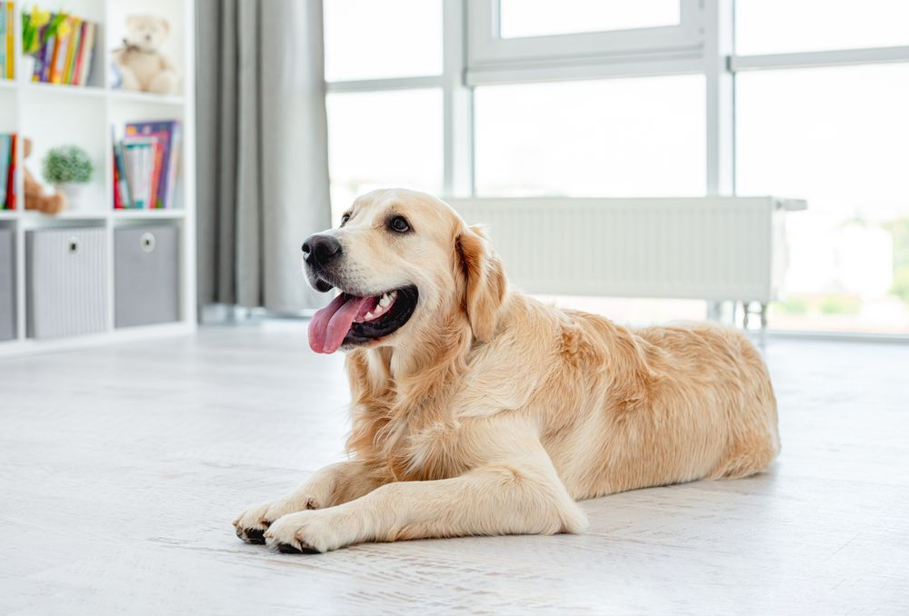
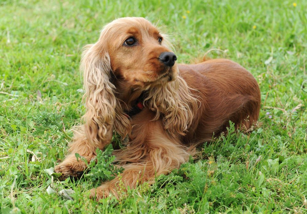
• What is the dog’s signalment? Various studies have shown that Cocker Spaniels, Poodles, Pyrenean Shepherds and Labrador Retrievers are all predisposed to developing otitis due to the conformation of their pinnae, EECs and/or hereditary susceptibility [5]. In young dogs, otitis can be caused by Otodectes cynotis – although this is less common with the newer oral and spot-on ectoparasiticides – whereas in old dogs an underlying endocrinopathy is more likely.
• What are the owner’s concerns? They may describe head shaking, ear rubbing, aural discharge and malodor [6] .
• When was the complaint first noticed? Abrupt and frenzied head shaking increases suspicion of an aural foreign body [6] , whereas chronic cases are usually associated with clinical or subclinical disease.
• Is the otitis unilateral or bilateral? Acute unilateral otitis increases the likelihood of an aural foreign body; chronic bilateral otitis is more likely to indicate other etiologies (e.g., allergies) and can additionally be complicated by the ear anatomy.
• What is the pet’s lifestyle? Does the dog exercise in fields or go swimming? Water trapped in the EEC changes the aural environment and can cause dysbiosis [6].
• Does the dog suffer from seasonal flares of otitis? If so, this is highly suggestive of primary allergic skin disease, such as non-food-induced atopic dermatitis.
• Has any previous topical treatment been successful? If not, this could indicate either a resistant infection or an adverse drug reaction.
Clinical examination
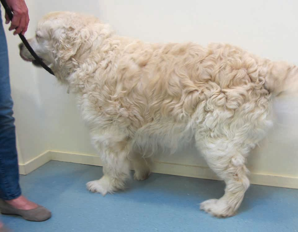
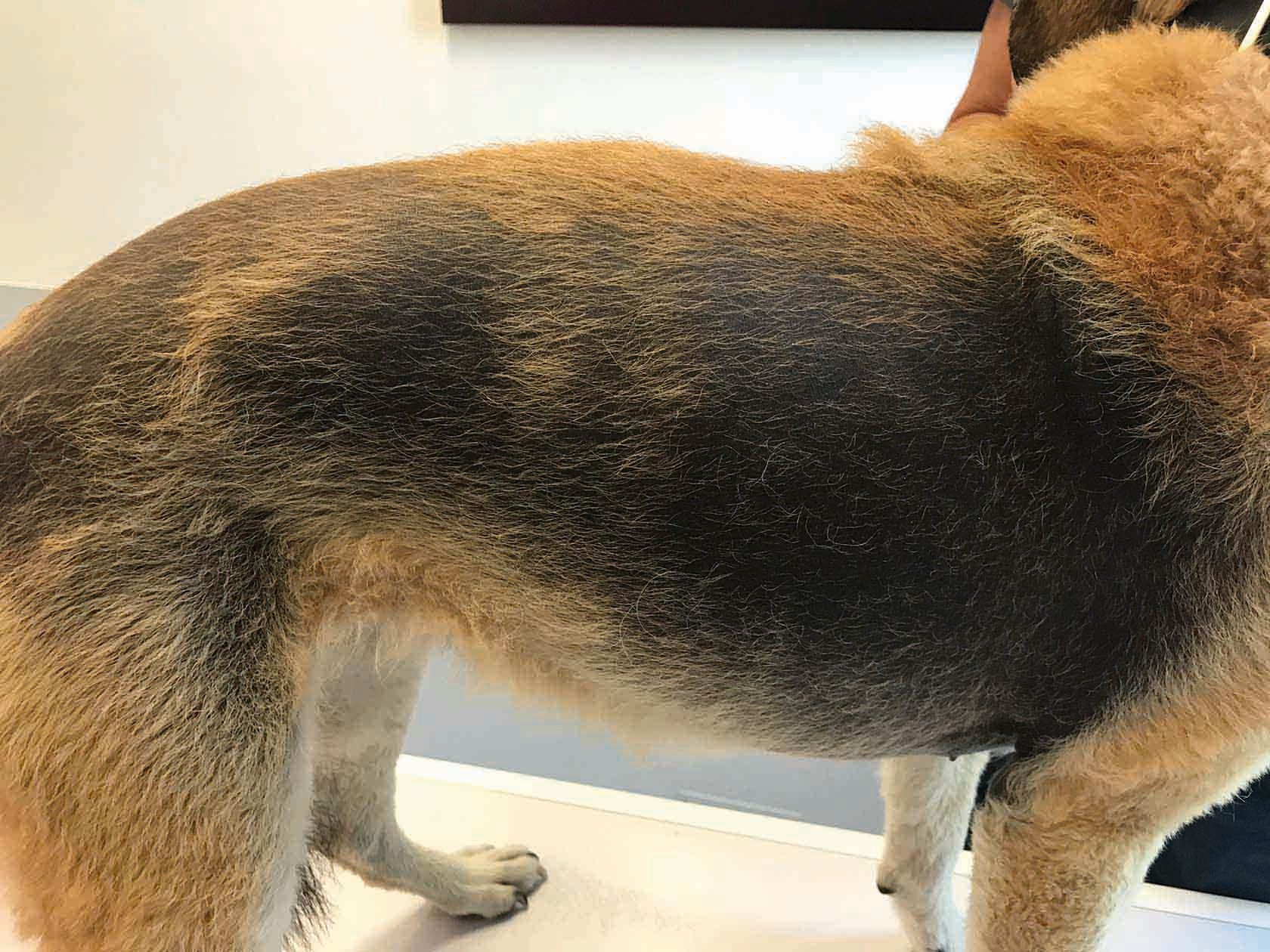
The next step is to perform a complete physical examination, followed by a specific dermatological examination. Clinicians typically have a routine for this, but generally it is advisable to start from the nose and work back towards the tail, thus ensuring all body systems are checked. When dealing with otitis, the physical examination may allow a tentative diagnose of OM, otitis interna (OI) or hypothyroidism. Clinical signs of OM include facial nerve paralysis (e.g., head tilt, ear droop, lip droop and ptosis) and Horner’s syndrome (i.e., miosis, ptosis, enophthalmos and protrusion of the nictitating membrane). Clinical signs of OI include hearing loss and vestibular disease (e.g., head tilt, asymmetric ataxia, leaning to the affected side, circling and horizontal nystagmus) [7 ], [8] . Hypothyroidism (other than the appearance of the skin and haircoat) is clinically associated with obesity, weakness, lethargy and bradycardia [9] . However, any suspect diagnosis should be confirmed with appropriate investigation(s).
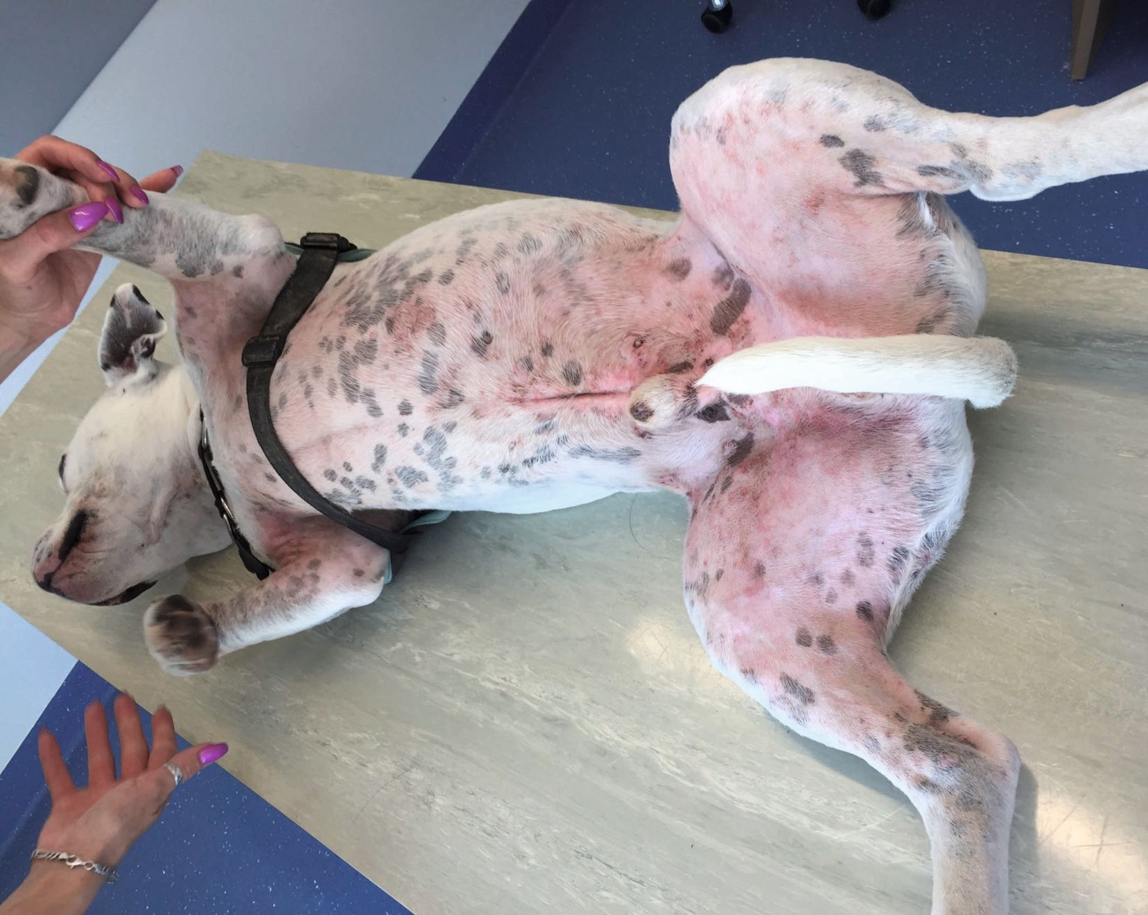
The dermatological examination should assess the skin in its entirety: periocular, perioral, dorsal and ventral neck, axillae, trunk (dorsum, ventrum and flanks), inguinal, perianal, interdigital (dorsal and palmar/plantar), pinnae and the EEC opening. Practitioners should be mindful of any skin lesions that could relate to otitis which may explain the primary etiology. For example, as well as signs of otitis, puppies with juvenile cellulitis may have erythema, oedema, exudation, crusting and alopecia of the face and muzzle [10] , and dogs with atopic dermatitis may present with a classic combination of otitis, pododermatitis and superficial pyoderma.
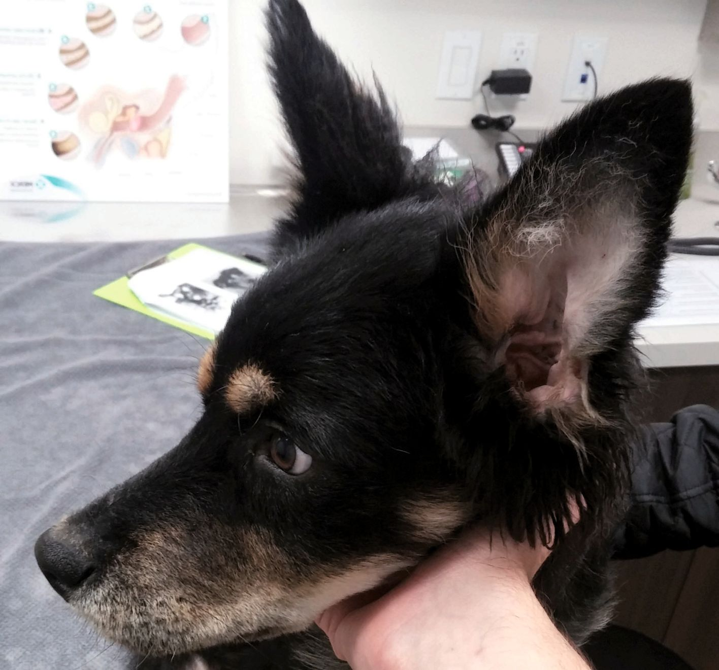
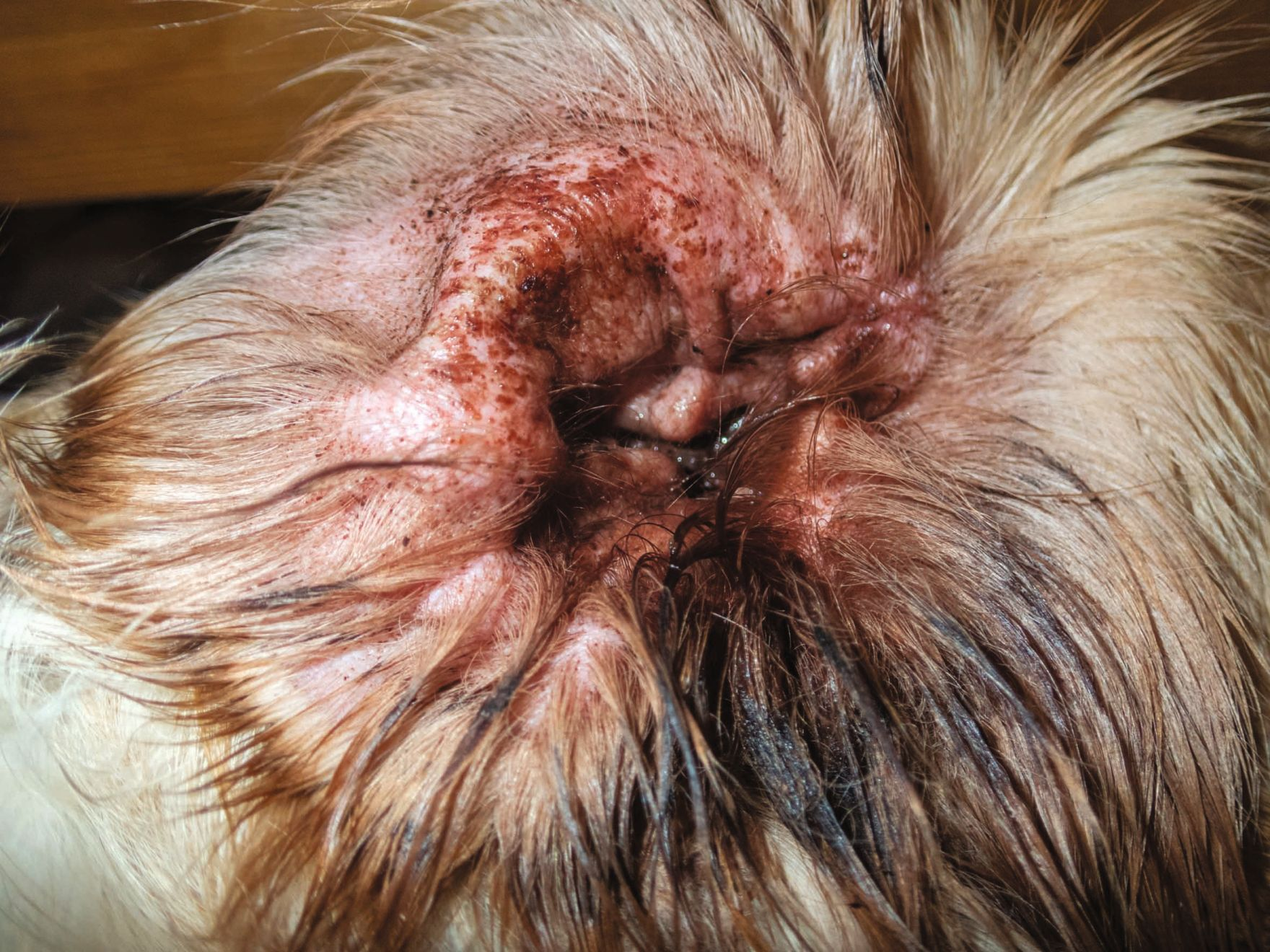
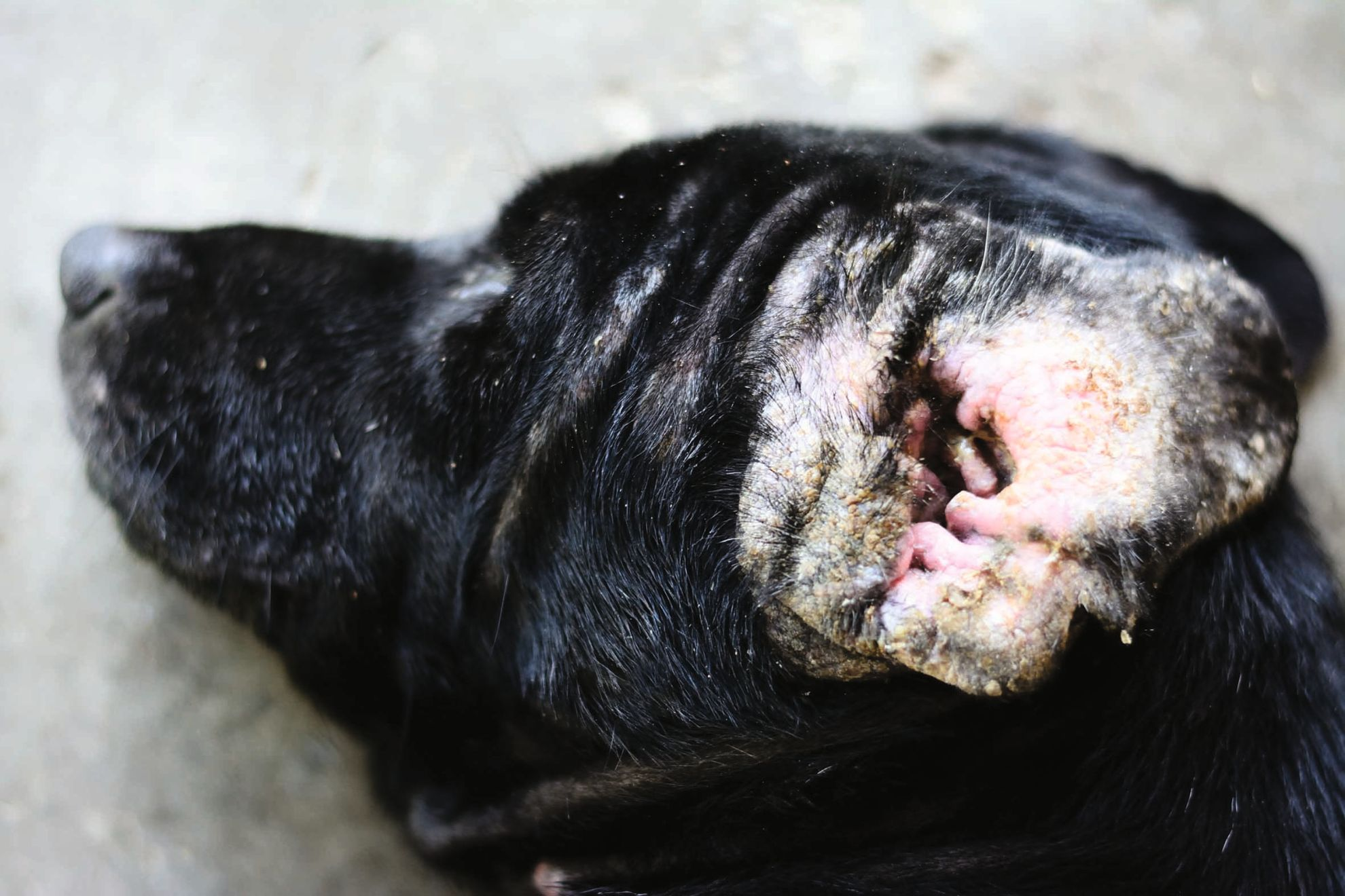
When dealing with otitis, it is sensible to carefully examine the ears last, as they can be painful, and dogs may subsequently develop an aversion to their ears being touched. However, even with minimal handling it is possible to collect more information simply by examining the inner aspect of the pinnae and opening to the EEC: erythematous pinnae can suggest an allergic etiology, whilst chronic cases may have thickened, hyperpigmented pinnae with excessive scaling, which may represent a cornification disorder [6] . Additionally, the appearance of any aural discharge can reveal primary or secondary causes for the otitis: a dry, brown, granular discharge is seen with O. cynotis, a moist, brown discharge commonly occurs with both staphylococcal and Malassezia infections (Figure 1), and a purulent, malodourous discharge is typical of Gram-negative bacterial infection (Figure 2) [2].
Otoscopy
If tolerated by the patient and once distant examination of the pinnae is complete, it is essential to perform otoscopy to evaluate the EEC and the integrity of the TM. There are three different types of otoscope available [11, ][12]:
1. Closed otoscope – allows good visualization of the EEC and TM and is designed to allow air to be introduced into the canal in order to undertake tympanometry, although accessing the EEC via the scope (e.g., to perform cytology) is limited.
2. Open otoscope – gives an inferior view of the EEC and TM compared to closed otoscopy but permits excellent access into the EEC. For this reason, all practices should have the option of an open otoscope.
3. Video otoscope – enables an excellent view and access to the EEC and TM, with the extra benefit of taking photographs and videos, although the equipment cost and the skill required for proper use can be an issue.
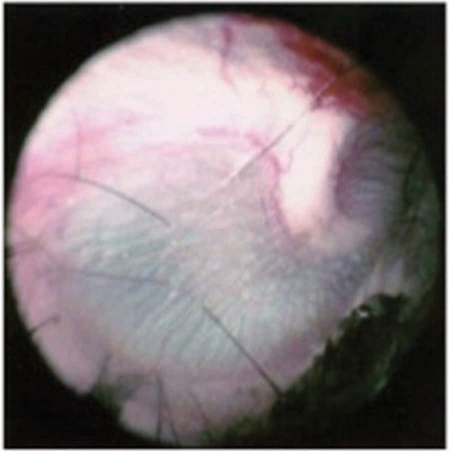
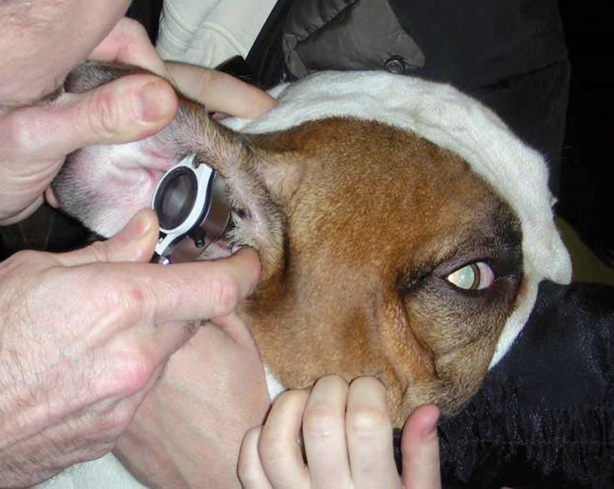
To fully appreciate the usefulness of otoscopy practitioners should be familiar with the appearance of the healthy aural anatomy. The normal EEC is a smooth, pale pink, thin-walled structure and the normal TM is a semi-transparent, concave membrane with a fine center and thicker periphery. The TM is divided anatomically into two: the dorsal section (pars flaccida) is light pink in color, whereas the ventral section (pars tensa) is pearl-grey (Figure 3). For every patient and each ear an appropriately sized, sterile otoscope cone (stored at room temperature) should be used. The cone will gently slide along the intertragic incisure – the soft depression separating the tragus and antitragus cartilages at the base of the pinna – and into the EEC. Assuming this is tolerated by the patient, the vertical and horizontal portions of the canal can be examined; the junction between the two sections is distinguished by a prominent cartilaginous ridge, and the pinna should be elevated upwards and outwards to straighten the canal as best as possible [12]. The otoscope cone can then be eased into the horizontal portion for improved visualization (Figure 4). With experience it is possible to rapidly identify or assess for foreign bodies, O. cynotis, inflammation, exudate, stenosis, proliferation and TM status [11, ][12]. As with all the diagnostic steps performed so far, otoscopy also contributes to the etiological understanding of otitis (Table 1) [11,][12].
| Otoscopic finding(s) | Direct deduction |
|---|---|
| Erythematous and hyperplastic EEC | Acute otitis |
| Fibrotic and hard EEC | Chronic otitis |
| Erythema of the vertical ear canal with no discharge | Allergic otitis: primary etiology could be food-induced atopic dermatitis or non-food-induced atopic dermatitis |
| Erosions and ulcers of EEC with purulent discharge | Gram-negative bacterial infection |
| “Cobblestone” appearance of EEC lining | Sebaceous and ceruminous gland hyperplasia, capable of transitioning into polyp-like growths |
| Foreign bodies | Primary cause |
| Ectoparasites | Primary cause |
| Tumor | Predisposing factor |
Microscopic evaluation
After otoscopy it is essential to take a swab from the affected ear(s) for in-house cytology, and this should be performed for every patient. Obtaining a sample simply involves placing a cotton bud into the EEC for a few seconds, but the material in the horizontal canal is usually of most clinical relevance and safely swabbing this portion in conscious patients can be difficult. Therefore, advancing the bud until the cartilaginous ridge is reached, and swabbing at this junction, should be sufficient. The bud is then rolled onto a clean microscope slide and labelled [2]. Separate slides are prepared for cytology and to check for ectoparasites (e.g., O. cynotis and Demodex canis), especially if the patient is a young dog. The ectoparasite slide should be prepared with a few drops of mineral oil, the sample rolled on top and finished with a coverslip. Microscopic visualization for ectoparasites is maximized by using a low power (x4 or x10) objective and light intensity, and a closed condenser. The entire slide should be examined methodically with a “back and forth” or “up and down” pattern [2].The slide for in-house cytology should be stained using a commercial modified Wright kit, consisting of a fixative and eosin and hematoxylin stains. The slide should be dipped for approximately 5 seconds, rinsed and dried. Starting on low microscopic power (x4 objective) and light intensity, and with an open condenser, a cellular area on the slide should be focused on. The power should then be increased to the highest power (x100 oil immersion objective), which will allow micro-organisms and inflammatory cells to be identified [2, ][14].
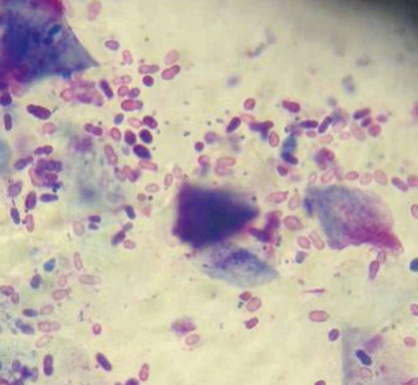
Under normal circumstances low numbers of bacteria (e.g., coagulase-negative Staphylococcus spp., coagulase-positive Staphylococcus spp. and Streptococcus spp.) and yeasts (predominantly Malassezia pachydermatis) reside in the canine EEC. When the canal becomes insulted or inflamed, bacteria and/or yeasts can become opportunistic, overgrow and potentially cause infection. Studies have suggested mean micro-organism numbers per high power field (x40 objective) indicative of normal microflora versus an abnormally increased population; for bacteria this is 5 or less versus 25 or more, and for Malassezia it is 2 or less versus 5 or more (Figure 5). Moreover, and in contrast to the normal aural flora, micro-organisms routinely contributing to otitis are coagulase-positive staphylococci, β-hemolytic streptococci, Pseudomonas spp. and Proteus spp. [2, ][15].
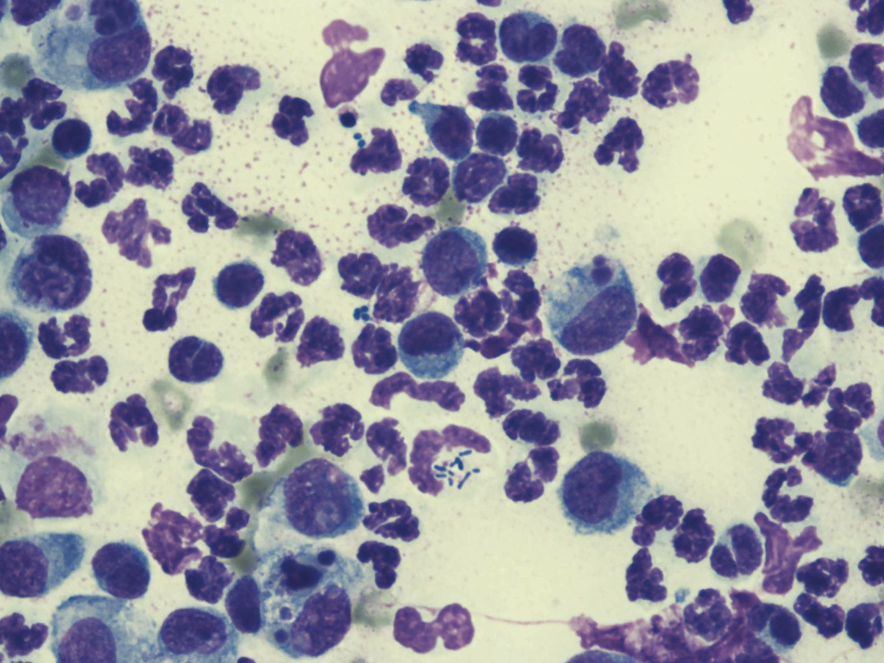
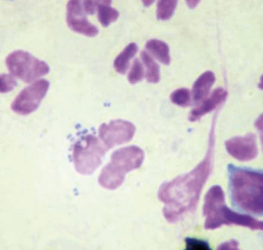
Cytology also helps determine overgrowth from infection by the presence of inflammatory cells [predominantly degenerate or non-degenerate neutrophils (Figure 6)] but it is not possible to identify bacterial species on cytology – culture is required for this. Culture and sensitivity (C&S) testing should not be used routinely, but rather reserved for certain scenarios, as culture cannot distinguish between resident bacteria, overgrowth and infection, so the reported antibiotic sensitivity is for all micro-organisms present. The reporting of irrelevant bacteria can lead to inappropriate antibiotic therapy or unnecessary switching of treatment. Conversely, C&S may fail to culture important micro-organisms, misleading interpretation and causing a premature cessation of treatment [2]. Culture is certainly indicated for chronic and medically unresponsive cases of OE, when rod-shaped bacteria are identified on cytology, or if OM is present. Additionally, studies have proven that different micro-organisms can independently cause infections in the external and middle ear; patients with OE and OM should therefore have samples taken from both areas, which could potentially produce two sets of results with differing antibiotic sensitivity patterns [16].
Otoscopic examination may be challenged by the patient’s anatomy, pathology and temperament; if encountered, it is preferable to perform otoscopy under sedation or general anesthesia and, where stenosis is present, after a course of oral glucocorticoids.
Diagnostic imaging
Imaging can allow further assessment of an otitis case, and especially the status of the middle ear. The literature recommends imaging for cases of suspected OM, para-aural abscessation, trauma, nasopharyngeal polyps, neurological dysfunction, and if a dog is unable to open its mouth 17. Moreover, imaging can help determine the direction of treatment: medical versus surgical. Ear canals that are associated with bony and irreversible pathological changes are more likely to be treated surgically 18.
• Radiography of the skull to assess the EEC and middle ear should be done under GA; this should consist of left and right oblique views, a dorsoventral skull view and an open-mouth rostrocaudal view; the latter is the best option to evaluate the tympanic bulla (TB). Imaging may confirm occlusion and bony changes of the EEC, content within the TB, and lysis or proliferation of the TB wall. However, the pathology must be severe to be detectable, and subtle changes are easily missed 1719. Radiography can also be used to assess the TM integrity using a technique called positive contrast canalography. This requires a soluble non-ionic iodine contrast medium to be introduced into the EEC and allowed to diffuse by gravity for a few minutes before obtaining dorsoventral and rostrocaudal open mouth radiographs. If the TM is ruptured, contrast medium may be identified in the middle ear; however, if a patient has stenotic ear canals the contrast may not reach as far as the middle ear, even if the TM is not intact. Consequently, caution must be used when interpreting the radiograph when using this procedure 20.
• Sonography allows the TB to be assessed, with the probe placed on the ventrolateral surface of each bulla; tiny movements of the probe allow the bullae to be scanned for fluid or a mass lesion. The major disadvantage of this modality is that a high level of skill is needed 171921.
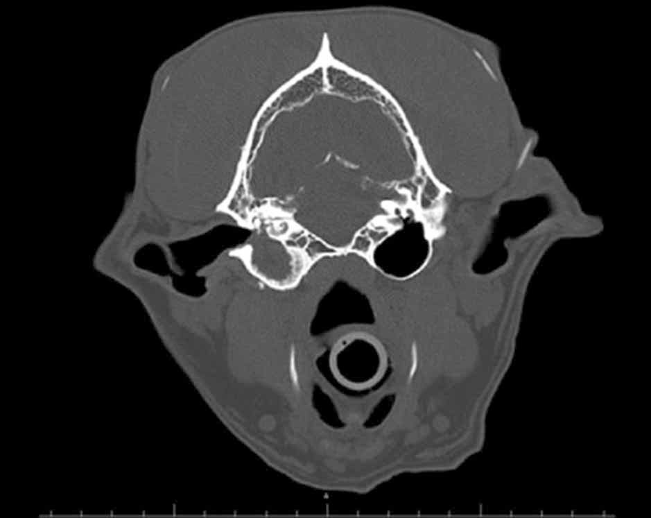
• Computed tomography (CT) and magnetic resonance imaging (MRI) are advanced imaging modalities which can be useful with some otitis cases. CT allows excellent visualization of the bony structures local to the ear, and is excellent for diagnosing stenosis or occlusion of the EEC and filling of the TB (Figure 7). MRI offers the best resolution for soft tissue structures, and is preferable if masses within or around the ear are suspected, although it is less sensitive at highlighting the cartilage of the EEC and the TB 1719.
Myringotomy
The TM will be intact in approximately 70% of OM cases, as the middle ear can become infected without OE by micro-organisms migrating from the pharynx via the auditory (Eustachian) tube or by hematogenous spread. Cavalier King Charles Spaniels and brachycephalic breeds can also have a primary OM with no EEC pathology [22]. In cases when OM is diagnosed but the TM is intact, myringotomy (iatrogenic rupture of the TM) is required. This is performed under GA guided by video otoscopy after the EEC has been thoroughly lavaged and allowed to dry. With direct visualization a 6F urinary catheter, cut obliquely at 60° and attached to a 2 mL syringe, is advanced through the most ventral part (6-7 o’clock) of the TM. One milliliter of sterile saline is infused into the middle ear and aspirated; the sample is transferred to a sterile tube and centrifuged to prepare samples for in-house cytology and C&S testing. If the middle ear subsequently requires further treatment the puncture site can be carefully enlarged for easier access and repeatedly lavaged until clean and empty [7,] [11]
Conclusion
Practitioners should follow a step-by-step approach when dealing with canine otitis to avoid misunderstanding the case, as this will inevitably lead to treatment failure. It is important to consider both primary and secondary causes, as well as predisposing and perpetuating factors, and a logical progression will elicit useful information that helps confirm the status of both the external canal and the middle ear. In short, the more thorough the investigation, the more likely is long-term treatment success.

Hannah Lipscomb
United Kingdom
Dr. Lipscomb gained her degree from London’s Royal Veterinary College in 2016 and was the first dermatology intern at Southern Counties Veterinary Specialists (SCVS), working alongside the specialist Filippo De Bellis. After qualification she initially worked in a first opinion small animal practice for 18 months – where she developed her interest in dermatology – and then progressed to a rotating internship at a private referral clinic before moving to SCVS to undertake a dermatology internship.
Filippo De Bellis
DVM, CertVD, Dip. ECVD, MRCVS
United Kingdom
Dr. De Bellis qualified from the University of Bari, Italy in 2001 and moved to the UK to undertake a dermatology residency at the Royal Veterinary College in 2006. He gained the RCVS Certificate in Veterinary Dermatology in 2009 and a year later became a Diplomate of the European College of Veterinary Dermatology. He has a particular interest in ear diseases and allergies, and is currently Head of Dermatology Services at Davies Veterinary Specialists, Southfields Veterinary Specialists and London Vet Specialists.
References
- Hill PB, Lo A, Eden CAN, et al.Survey of the prevalence, diagnosis and treatment of dermatological conditions in small animals in general practice. Vet Rec 2006;158(16):533-539.
- Angus JC. Otic cytology in health and disease. Vet Clin North Am Small Anim Pract 2004;34(2): 411-424.
- Shell LG. Otitis media and otitis interna; etiology, diagnosis and medical management. Vet Clin North Am Small Animal Pract 1988;18(4):885-899.
- Bajwa J. Canine otitis externa – treatment and complications. Can Vet J 2019;60(1):97-99.
- Saridomichelakis MN, Farmaki R, Leontides LS et al. Aetiology of canine otitis externa: a retrospective study of 100 cases. Vet Dermatol 2007;18(5):341-347.
- August JR. Otitis externa. Vet Clin North Am Small Anim Pract 1988;18(4):731-742.
- Gotthelf LN. Diagnosis and treatment of otitis media in dogs and cats. Vet Clin North Am Small Anim Pract 2004;34(2):469-487.
- Cook LB. Neurological evaluation of the ear. Vet Clin North Am Small Anim Pract 2004;34(2): 425-435.
- Panciera DL. Hypothyroidism in dogs: 66 cases (1987-1992). J Am Vet Med Assoc 1994;204 (5):761-767.
- Miller Jr WH, Griffin CE, Campbell KL. Miscellaneous Skin Diseases. In: Saunders (ed). Muller and Kirk’s Small Animal Dermatology. 7th ed. Missouri: Elsevier Mosby, 2013;708-709.
- Griffin CE. Otitis techniques to improve practice. Clin Tech Small Anim Pract 2006;21(3):96-105.
- Cole LK. Otoscopic evaluation of the ear canal. Vet Clin North Am Small Anim Pract 2004;34(2):397-410..
- Little CJ, Lane JG. An evaluation of tympanometry, otoscopy and palpation for assessment of the canine tympanic membrane. Vet Rec 1989;124(1):5-8.
- Neuber A, Nuttall T. Introduction to Dermatological Tests. In: Neuber A, Nuttall T (eds). Diagnostic Techniques in Veterinary Dermatology. 1st ed. New Jersey: Wiley-Blackwell, 2017;10-12.
- Rosser Jr EJ. Causes of otitis externa. Vet Clin North Am Small Anim Pract 2004;34(2):459-468.
- Cole LK, Kwochka KW, Kowalski JJ, et al. Microbial flora and antimicrobial susceptibility patterns of isolated pathogens from the horizontal ear canal and middle ear in dogs with otitis media. J Am Vet Med Assoc 1998;212(4):534-538.
- Benigni L, Lamb C. Diagnostic imaging of the ear disease in the dog and cat. In Pract 2006;28(3):122-130.
- Doyle RS, Skelly C, Bellenger CR. Surgical management of 43 cases of chronic otitis externa in the dog. Ir Vet J 2004;57(1):22-30.
- Garosi LS, Dennis R, Schwarz T. Review of diagnostic imaging of ear diseases in the dog and cat. Vet Radiol Ultrasound 2003;44(2):137-146.
- Trower ND, Gregory SP, Renfrew H, et al. Evaluation of the canine tympanic membrane by positive contrast ear canalography. Vet Rec 1998;142(4):78-81.
- Dickie AM, Doust R, Cromarty L, et al. Ultrasound imaging of the canine tympanic bulla. Res Vet Sci 2003;75(2):121-126.
- Milne E, Nuttall T, Marioni-Henry, et al. Cytological and microbiological characteristics of middle ear effusions in brachycephalic dogs. J Vet Intern Med 2020;34(4):1454-1463.